> Biology Group. 2021 Feb 26;2(2):084-090. doi: 10.37871/jbres1193.
Cytogenetic Study on Mitotic Cell Division in Allium cepa by Lead (Pb) and Chromium (Cr) Containing Bacterial Strain Isolated from Tannery Effluents of Bangladesh
Afrin Priya Talukder, Sarwat Tazrian, Md. Nazmul Haque, Shahriar Zaman and Md. Akhtar-E-Ekram*
- Lead and chromium
- 16S rRNA sequencing
- Myroides sp
- Cytogenetic effect
- Allium cepa
- Cytotoxicity test
In the present study, a bacterial strain capable of Pb and Cr detoxification was isolated from tannery effluents which was identified as Myroides sp. through 16S rRNA gene sequencing. In the cytogenetic experiment, 100 and 600 µg/ml of lead and chromium were used as treatment for the root tips of Allium cepa and caused many chromosomal abnormalities such as abnormal chromosome position, damaged nucleus, breaks of chromosome bridges and fragments also occurred. Notably, sticky metaphase was found where sticky chromosomes indicated highly toxic, usually irreversible consequences leading to cell death. However, Myroides sp. treated supernatant, collected after day 7, used to treat Allium cepa tips showed less mitotic aberrations, nuclear degeneration and observed normal anaphase and telophase stage indicating possible metal detoxifying ability of the isolated strain. Furthermore, LC50 value was 64.63 μl/ml for Myroides sp.
Heavy metals are the most recurrent inorganic pollutants in water [1] and are highly distributed over the earth’s crust [2]. Epidemiological evidence suggested that a long-term exposure to heavy metals was highly associated with increased risk of development of several diseases including cancers [3]. In vivo and in vitro assays have shown that heavy metals induce chromosomal aberrations and micronucleus in plants [4]. Lead and chromium are considered to be of highest concern because of their genotoxicity in microorganisms, animals and humans. Acute lead poisoning results in a well characterized syndrome manifested in adults by colic, anemia, headache, fatigue, gum line and peripheral neuropathy [5] and has also adverse effects on oxidative stress [6]. Moreover, toxicity of chromium has been associated with cancer and disruption to cellular functions [7]. It was reported that chromium concentrations should not be exceeded 80 µg/ml in sea water and 520 µg/ml in rivers and lakes [8]. Thus, our present investigation was planned to determine bacterial detoxifying ability at 100 µg/ml and 600 µg/ml concentrations of Pb and Cr salt, separately.
Genotoxicity identifies compounds present in the environment having the potential to cause mutation by damaging the DNA which can result into point mutation, change in the DNA structure or damage on the chromosome. Sometimes genotoxic effects such as deletions, breaks or rearrangements can lead to cancer if the damage does not immediately lead to cell death. Monitoring of genotoxic compounds in the environment has become an important issue for public health, with the purpose of minimizing direct and indirect human exposure to these toxic substances. In the search of pertinent short term test systems, plant materials have proved to be useful in basic research of toxicity bioassay as well as part of a test battery in environmental monitoring. Plant assays are quite easy to accomplish, low-priced, rapid and good predictors of genotoxicity [9]. Bioassays with plants such as Allium cepa test is important since it is an excellent model in vivo, where the roots are in direct contact with the substance of interest (i.e. effluents that being tested) enabling possible damage to the DNA of the eukaryotes to be envisaged. Recent studies have shown that, several bacteria strains are able to detoxify these heavy metals, those which are harmful for plants as well as for humans and animals [10]. In this study, by using metal detoxifying isolate, the reduction of the toxic effect of lead and chromium were examined through cell division in the response of Allium cepa.
Sample collection
The waste water sample was collected from the discharge and drainage pipes of leather industrial zone at Hazaribagh, Dhaka, Bangladesh, during January 2016.
Isolation of bacterium
The collected tannery effluent was filtered by filter paper in a beaker and 5 ml of the tannery effluents were added to 100ml MS medium separately contained lead as Lead Nitrate [Pb(NO3)2] and chromium as Chromium Nitrate [Cr(NO3)3] at different concentrations (100µg/ml, 200µg/ml, 300µg/ml, 400µg/ml, 500µg/ml, 600µg/ml) for enrichment and selection. Pb and Cr degrading bacterium was isolated by plating from the old bacterial suspension of the liquid medium into an agar solidified MS medium by the standard pour plate method supplemented with different concentrations of lead and chromium. The plates were then incubated at 37°C for 5 days and bacterial colonies were found to grow on the medium. Isolated bacterial colony was purified by re-streaking further on nutrient agar medium and incubated for 18-24 hour at 37°C. Then the isolate was preserved in 30% glycerol at -20°C.
Molecular identification of isolated bacterium
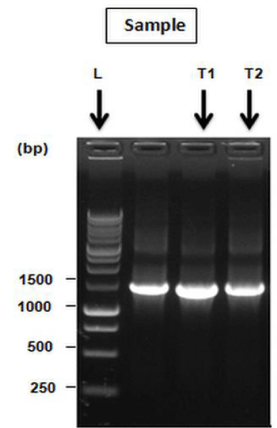
16S rRNA identification is considered the most reliable tools for identification of microorganism species [11]. The chromosomal DNA was extracted from the bacterial culture and the extracted DNA was used as template for PCR to amplify 16S rRNA gene. 8F(5`AGA GTT TGA TCM TGG CTC AG3`) and 1492R(5`GGT TAC CTT GTT ACG ACT T3`) were used as forward and reverse primer, respectively. After PCR amplification the gel was run and desired band was found. Next the PCR product was purified and the concentration of purified product was measured using the Nano Drop ND-2000 Spectrophotometer and diluted with sterile water to 50 ng/ml before samples were sequenced. Sequencing was done from both forward and reverse ends using either 8F or 1492R primers and carried out in ABI Prisom 310 machine. 16S rRNA gene sequence of selected bacterial isolate was compared with other reference sequences as available in the NCBI database using the Basic Alignment Search Tool (BLAST) algorithm.
Cytogenetic study
The test material used was onion bulbs from a population of commercial varieties of Allium cepa. At first, commercially available onions were soaked into distilled water for 2 or 3 hours, were put on the test tube, filled with distilled water and kept in a dark place at room temperature for growing roots and after 48 hours of exposure, Allium cepa roots were observed. The most important macroscopic parameter was the root growth. When Allium cepa roots were observed after 48 hours, the length of the root bundle was measured using a calibrated ruler in mm. Three longest roots in each bulb were selected and used for the experiment out of ten onions. Here, the isolated strain was used as test sample to determine the detoxifying capability of the bacterium towards lead and chromium. Before each test, bacterial cultures were centrifuged and 10 ml supernatant was then diluted with 10 ml water which was used as the treatment solution supplemented with different concentrations (100 µg/ml and 600 µg/ml) of Pb and Cr up to 7 days. After 48 hours, the onions were set up for 24 hours in each test solution. At the same time, some of the onions were grown in control medium (Distilled water) for 24 hours.
From both of control and treatment medium roots were collected by a pair of fine forceps and were fixed in aceto-alcohol (1:3) solution. After 24 hours of fixation, root tips were transferred from fixative to 70% ethanol and stored in the refrigerator until they were used in the laboratory. After removing from 70% ethanol the root tips were washed with distilled water for 10 minutes. Washed root tips were then transferred to 1N HCL for 7 minutes and the root tips were washed again with distilled water for 5 minutes. The mordanting was done with 2% aqueous solution of iron alum (Ferric ammonium sulfate) for 7 minutes. The root tips were washed again with frequent changes of distilled water for 7 minutes in order to complete removal of the mordanting fluid from the tissues. Then root tips were stained with 0.5% hematoxylin for 10 minutes. Finally, root tips were washed with distilled water for 5 minutes. The stained root tip was then taken on a clean slide and meristamatic zone (1-1.5 mm portion) of it was cut with a razor blade and a drop of 0.5% acetocarmine was added. The cells were then covered with a cover glass and slight pressure was applied by a pencil eraser over the cover glass. The slide was warmed over an alcohol flame and again a slight pressure was applied by finger tips over the cover glass keeping the slide duly wrapped in blotting paper. All slides were marked and examined under a microscope (LABOMED CXL, USA) with 40X magnification. Then, mitotic abnormalities in treatment medium were recorded and compared to control.
Cytotoxicity test of isolated bacterium culture
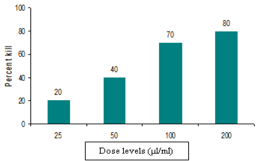
In our present study, toxicity test of bacterial supernatant was performed through brine shrimp lethality bioassay to make confirmation about bacterial toxic protein secretion and its impacts on disrupting cellular functions. Brine shrimp lethality bioassay is considered a useful tool for preliminary assessment of toxicity [12]. Brine shrimp eggs were hatched in simulated seawater to get nauplii. For test sample, 10 ml of bacterial culture were taken in 10 eppendorf tubes and were centrifuged at 7000 rpm for 10 min. Supernatants were collected which was further used as test sample and specific volumes of test sample were transferred to the respective vials to get, final concentrations of 25 µl/ml, 50 µl/ml, 75 µl/ml, 100 µl/ml, 125 µl/ml, 150 µl/ml, 175 µl/ml and 200 µl/ml, respectively. Survivors were counted after 24 hours and these data were processed in a simple program for probit analysis to estimate LC50 value with 95% confidence intervals.
Molecular identification of isolated bacterium
After gene sequencing, the sequence was checked against the 16S rRNA gene sequences of other organisms that had already been submitted to NCBI Gene bank database. Isolated bacterium showed 98% identity with Myroides sp. (Accession no. MG733977). The band of PCR product of isolated bacterium was shown in figure 1.
Cytogenetic study on Allium cepa
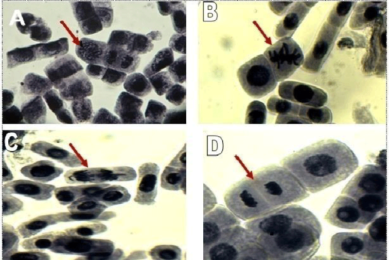
Control samples indicated normal mitotic cell division. The figure 2(A) clearly showed the prophase stage in which the chromatin shortens and thickens into double stranded chromosomes and the nuclear membrane disintegrates. Normal metaphase is shown in figure 2(B) where the spindle fibers align as double stranded chromosomes in single file along the equator of the cell, metaphase plate. In anaphase (Figure 2C), the spindle fibers pull on the centromeres of the chromosomes separating the sister chromatids of each double stranded chromosome. Each single strand is pulled to an opposite pole of the cell. Consequently in telophase (Figure 2D) the chromatids reach the opposite poles of the cell and the spindle detaches and new nuclear membranes form around the chromatids where cytokinesis begins even before telophase completes with the formation of a cleavage furrow (indentation of the cell membrane). In control experiment, 30% dividing cells (Prophase-17.5%, Metaphase-4.5%, Anaphase-3%, Telophase-5.5%) were found among 2000 cells.
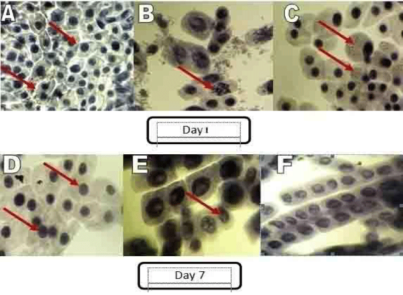
The cyto morphological study indicated adverse effects of chemical on the root tip cells compared to the control untreated group (Figure 3). Low dose of lead (100 µg/ml) on day 1 caused the appearance of many chromosomal abnormalities (Figure 3A) as reported earlier [13]. Lead also caused development of extensive vacuoles in the root tip cells growing in 100 µg/ml (Figure 3C). The vacuoles of root tip cells are the major sites of metal sequestration under chronic exposure scenario and it is thought to be a detoxification pathway for preventing cell damage by retaining metals in specific vacuoles [14]. Allium cepa root tip cells in the highest dose (600 µg/ml) exposed group, after 24 hours exhibited nuclear disruption and condensation of the chromatin material, indicating apoptotic death of root tips cells (Figure 3B). A similar finding of apoptotic death of root tips cells after aluminium exposure was reported in barley [15]. Root tip of Allium cepa treated with supernatant, containing low dose of lead (100 µg/ml) that was treated with bacterium for 7 days, showed less mitotic aberration and absence of vacuoles (Figure 3D). High dose (600 µg/ml) of the lead sample also gave the similar result, decreasing the presence of nuclear condensation and abnormal positioning of the chromosome (Figures 3E, 3F). In case of treatment with bacterial supernatant containing lead, the percentage of dividing cells (Improper metaphase-7.5%, Sticky telophase-6%, Vacuolation-4%) were 17.5% among 2000 cells at day 1 of exposure and 25 % dividing cells (Less nuclear condensation-15%, Normal structure of cell-10%) were observed among 2000 cells at day 7 of exposure.
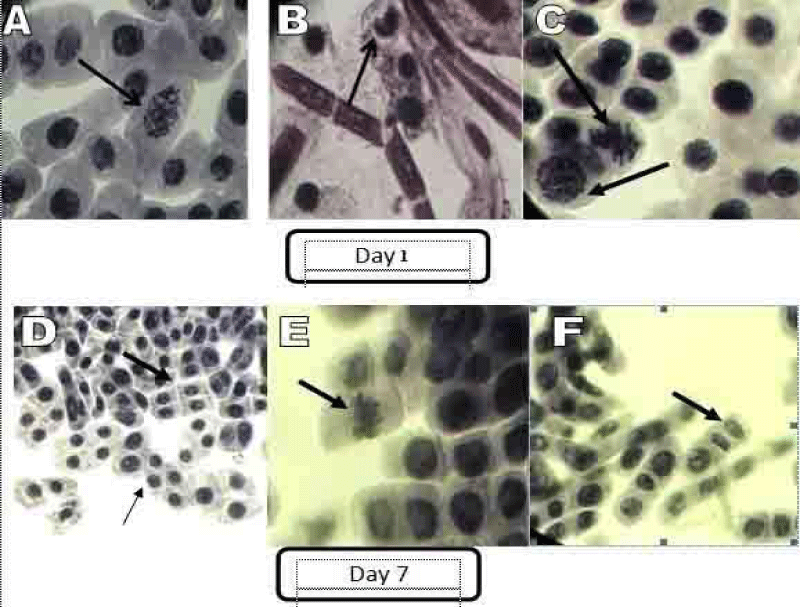
Low and high doses of chromium (100 and 600µg/ml) caused many chromosomal abnormalities from day 1. Chromosomes took abnormal positions dissimilar to normal phases of mitosis cell divisions while abnormal and degenerating nucleus indicated the toxicity of chromium (Figures 4A, 4B). Chromosome bridges and fragments resulted from chromosome and chromatid breaks occurred. Sticky metaphase was evident in figure 4(C). Sticky chromosomes indicated highly toxic, usually irreversible effects probably leading to cell death and the presence of chromosome adherences (stickiness) may be a sign of genotoxic effects. These aberrations may originate from either translocations or cohesive chromosome terminations. Bacteria treated supernatants at day 7 showed less mitotic aberrations and nuclear degeneration (Figure 4D). They also showed normal anaphase and telophase stage (Figures 4E, 4F). In case of treatment with bacterial supernatant containing chromium, 20% dividing cells (Nuclear degeneration-9%, Abnormal shape of nucleus-8%, Sticky metaphase-3% ) were found among 2000 cells at day 1 of exposure whereas 27.5 % dividing cells (Normal metaphase-9%, Normal telophase-7.5%, Less nuclear degeneration-11%) were observed among 2000 cells at day 7 of exposure.
Cytotoxicity test of isolated bacterium culture
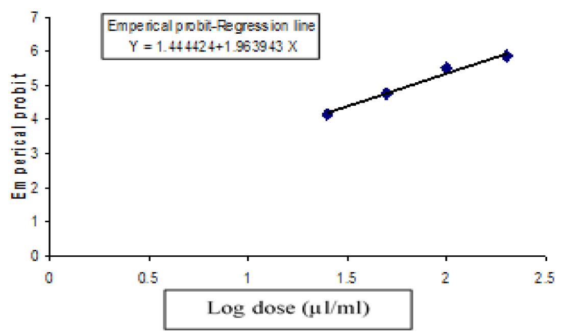
The cytotoxic effect of isolated bacterial supernatant was studied at concentrations of 25 µl/ml, 50 µl/ml, 75 µl/ml, 100 µl/ml, 125 µl/ml, 150 µl/ml, 175 µl/ml and 200 µl/ml. From the result, it was found that LC50 was 64.63 ± 0.13 µl/ml and the regression equation was, Y = 1.963x + 1.444, while the 95% confidence limits were 39.34 to 106.19 µl/ml for the 48 hours of exposure (Table 1, figures 5, 6).
| Table 1: Cytotoxic activity: LC50, 95% confidence limits, regression equations and Chi-square values for isolated bacterium culture against brine shrimp nauplii after 48 hrs of exposure. | ||||
| Test sample | LC50 (μl/ml) | 95% Confidence limits (μl/ml) | Regression equation | χ2 value (Degrees of freedom) |
| Isolated bacterium culture | 64.63 ± 0.13 | 39.34 -106.19 | Y = 1.963x + 1.444 | 0.154(2) |
Environmental pollution by heavy metals is always considered as a serious threat to living organisms in an ecosystem [16,17]. Industrial tannery wastewater is a major source of heavy metal contamination in our environment as heavy metal is of economic significance in industrial use [18]. Several approaches were undertaken to isolate and characterize heavy metal tolerant and degrading bacteria from industrial effluents [19]. Microorganisms with catabolic potential and their products such as enzymes and bio surfactant are directly used to enhance and boost their remediation efficacy [20]. The Myroides sp. are prevalent as environmental bacterial organisms [21]. Like other bacteria, Myroides sp. can adsorb metal ions by ionazable groups of the cell wall or capsule (Carboxyl, Amino, Phosphate and Hydroxyl groups) [22]. In the present investigation, lead and chromium detoxifying bacterial strain (Yellow colored colony) was isolated from tannery effluents and cultured on MS agar solidified medium supplemented with metals (Lead and chromium) by using streak plate and pour plate methods.
In the present study, sequencing was done by 16S rRNA gene for molecular identification of the isolated bacterial strain. Comparison of the bacterial 16S rRNA gene sequence has emerged as a preferred genetic technique [23]. Furthermore, 16S rRNA gene (1.5 kb in length) has emerged as a useful molecular tool since it is present in all bacteria, either as a single copy or in multiple copies as well as it is highly conserved over time within a species [24]. Sequencing result of 16S rRNA gene confirmed that the bacterial isolate showed 98% significant alignments with Myroides sp. Although Sanger sequencing of the 16S ribosomal RNA gene is used as a molecular method, identification of bacteria to the species level is not always possible due to high sequence similarities between some species [25]. Moreover, sequencing results are relative rather than absolute in which the actual quantity of a particular bacteria is uncertain. In addition, 16S rRNA gene sequencing could be a result of uncontrollable PCR biasness because of each 16S rRNA gene may not amplify with equal efficiency during PCR reactions due to differential primer affinity and GC content [26].
Generally, induction of structural and numerical changes in chromosomes is a common criterion for recognizing potential genetic hazards of a particular agent. Genotoxic and mutagenic action of heavy metals were evaluated using different cytogenetic techniques applied to the Allium cepa test organism [27]. According to a spectrum of morphological abnormalities such as bridge chromosome, improper metaphase, vacuolation and pyncnotic nuclei were observed in root meristems after exposure to lead [28]. Several in vivo and in vitro studies have also shown that chromium compounds damage DNA in a variety of ways, including DNA Single And Double-Strand Breaks (SDSBs) generating chromosomal aberrations, micronucleus formation, sister chromatid exchanges, formation of DNA adducts, and alteration in DNA replication and transcription [29]. According to studies carried out by Seth, et al. [30] microbial sequestration of heavy metals show significant improvement in bioremediation of these heavy metals. Abbas, et al. [31] reported the heavy metals (Pb, Cr, Cu, Fe, Mn) detoxifying (50%) ability of Pseudomonas sp. within 7 days. In our study we also found that, by treating Pb and Cr with this bacterial isolate for about 7 days significant changes are found in A. cepa root cells, including less occurrence of chromosomal aberrations and nuclear condensation. Our findings showed similarity with the referenced data and from the result we can consider that the high levels of heavy metals induce the genotoxic effects observed in root cells of A. cepa. At the same time, we tried to find out toxicity of Myroides sp. at cellular level on brine shrimp.
Cytotoxic compounds generally show significant activity in the brine shrimp bioassay and this assay can be recommended as a guide for the detection of toxic compounds because of its simplicity and low cost [32]. In our present study, the test was done to confirm whether the bacterium had any effect on mitotic abnormalities because if the bacterium showed toxicity it might secrete extracellular toxic proteins in supernatant. Bacterial protein secretion systems have been an important spotlight in case of bacterial pathogenesis and our detoxifying bacterium was a gram negative pathogenic strain. Gram negative bacteria secrete extracellular proteins and thereby utilize a multitude of methods to invade hosts, damage tissue sites and prevent from biologically responding [33]. In the present study, isolated bacterial supernatant showed less toxic effect against Artemia salina with 64.63 ± 0.13 µl/ml LC50 value after 48 hours of exposure and it also confirmed that there was no significant impact on mitotic cell division in case of genotoxic effects on Allium cepa.
The potential application of heavy metals detoxification in the forms of toxic solid and liquid wastes by microorganisms has several advantages. By isolating and using bacteria we can able to reduce the amount of metals from the contaminated water or soil and can reduce the effect of cellular damage that is occurred by the metal poisoning. The results from the present study indicated Pb and Cr detoxifying capability of Myroides sp. by reducing the mitotic abnormalities in root cells of Allium cepa which may affect the normal growth and development at field level. Therefore, the cytotoxic effect of isolated bacterial supernatant enunciated that it can be selected for further experimental assay as it had a little toxic effect at cellular level on Artemia salina.
We acknowledge Microbiology Laboratory, Dept. of Genetic Engineering & Biotechnology, University of Rajshahi for technical facilities.
- Chandra S, Chauhan LK, Murthy RC, Saxena PN, Pande PN, Gupta SK. Comparative biomonitoring of leachates from hazardous solid waste of two industries using Allium test. Sci Total Environ. 2005 Jul 15;347(1-3):46-52. doi: 10.1016/j.scitotenv.2005.01.002. PMID: 16084966.
- Arambašić MB, Bjelić S, Subakov G. Acute toxicity of heavy metals (copper, lead, zinc), phenol and sodium on Allium cepa L., Lepidium sativum L. and Daphnia magna St.: Comparative investigations and the practical applications. Wat Res. 1995 Feb; 29(2): 497-503. doi:10.1016/0043-1354(94)00178-A.
- Zhuang P, McBride MB, Xia H, Li N, Li Z. Health risk from heavy metals via consumption of food crops in the vicinity of Dabaoshan mine, South China. Sci Total Environ. 2009 Feb 15;407(5):1551-61. doi: 10.1016/j.scitotenv.2008.10.061. Epub 2008 Dec 9. PMID: 19068266.
- Rodriguez-Cea A, Ayllon F, Garcia-Vazquez E. Micronucleus test in freshwater fish species: an evaluation of its sensitivity for application in field surveys. Ecotoxicol Environ Saf. 2003 Nov;56(3):442-8. doi: 10.1016/s0147-6513(03)00073-3. PMID: 14575685.
- Singh V, Chauhan PK, Kanta R, Dhewa T, Kumar V. Isolation and characterization of Pseudomonas resistant to heavy metals contaminants. Int J Pharm Sci Rev Res. 2014;3(2):165-168.
- Roy A, Queirolo E, Peregalli F, Mañay N, Martínez G, Kordas K. Association of blood lead levels with urinary F₂-8α isoprostane and 8-hydroxy-2-deoxy-guanosine concentrations in first-grade Uruguayan children. Environ Res. 2015 Jul;140:127-35. doi: 10.1016/j.envres.2015.03.001. Epub 2015 Apr 4. PMID: 25863186; PMCID: PMC4492803.
- Shobana SN. Characterization of chromium bioremediation by Stenotrophomonas maltophilia SRS 05 isolated from tannery effluent. Int Res J Eng Technol. 2017 Feb;4(2):403-429.
- Jacobs JA, Testa SM. Overview of chromium(VI) in the environment: background and history. In: Guertin J, Jacobs JA and Avakian CP (eds.). Chromium (VI) Handbook, Boca Raton; Fl CRC Press:2005, pp. 1-22.
- Panda BB, Panda KK. Genotoxicity and mutagenicity of metals in plants. Physiol biochem of metal toxicity and tolerance in plant. 2002, pp. 395-414.
- Bruins MR, Kapil S, Oehme FW. Microbial resistance to metals in the environment. Ecotoxicol Environ Saf. 2000 Mar;45(3):198-207. doi: 10.1006/eesa.1999.1860. PMID: 10702338s.
- Shah SU. Importance of genotoxicity & S2A guidelines for genotoxicity testing for pharmaceuticals. IOSR J Pharm Bio Sci. 2012;1(2):43-54.
- Solis PN, Wright CW, Anderson MM, Gupta MP, Phillipson JD. A microwell cytotoxicity assay using Artemia salina (brine shrimp). Planta Med. 1993 Jun;59(3):250-2. doi: 10.1055/s-2006-959661. PMID: 8316592.
- Wierzbicka M. Lead in the apoplast of Allium cepa L. root tips-ultrastructural studies. Plant Sci. 1998 Apr 1; 133(1):105-119. doi: 10.1016/S0168-9452(98)00023-5.
- Einicker-Lamas M, Mezian GA, Fernandes TB, Silva FL, Guerra F, Miranda K, Attias M, Oliveira MM. Euglena gracilis as a model for the study of Cu2+ and Zn2+ toxicity and accumulation in eukaryotic cells. Environ Pollut. 2002;120(3):779-86. doi: 10.1016/s0269-7491(02)00170-7. PMID: 12442801.
- Pan J, Zhu M, Chen H. Aluminum-induced cell death in root-tip cells of barley. Environ Exp Bot. 2001 Aug;46(1):71-79. doi: 10.1016/s0098-8472(01)00083-1. PMID: 11378174.
- Siddiquee S, Rovina K, Azad SA, Naher L, Suryani S, Chaikaew P. Heavy metal contaminants removal from wastewater using the potential filamentous fungi biomass: A review. J Microbial Bioch Technol. 2015;7(6):384-395. doi:10.4172/1948-5948.1000243.
- Okolo NV, Olowolafe EA, Akawu I, Okoduwa SIR. Effects of industrial effluents on soil resources in Challawa industrial area, Kano, Nigeria. J Glob Ecol Environ. 2016 July;5(1):1-10.
- Igiri BE, Okoduwa SIR, Idoko GO, Akabuogu EP, Adeyi AO, Ejiogu IK. Toxicity and Bioremediation of Heavy Metals Contaminated Ecosystem from Tannery Wastewater: A Review. J Toxicol. 2018 Sep 27;2018:2568038. doi: 10.1155/2018/2568038. PMID: 30363677; PMCID: PMC6180975.
- Magesh N, Chandrasekar N, Soundranayagam JP. Delineation of groundwater potential zones in Theni district, Tamil Nadu, using remote sensing, GIS and MIF techniques. Geosci Frontiers. 2012 March;3(2):189-196. doi:10.1016/j.gsf.2011.10.007.
- Le TT, Son MH, Nam IH, Yoon H, Kang YG, Chang YS. Transformation of hexabromocyclododecane in contaminated soil in association with microbial diversity. J Hazard Mater. 2017 Mar 5;325:82-89. doi: 10.1016/j.jhazmat.2016.11.058. Epub 2016 Nov 21. PMID: 27915102.
- Holmes B, Snell JJ, Lapage SP. Flavobacterium odoratum: a species resistant to a wide range of antimicrobial agents. J Clin Pathol. 1979 Jan;32(1):73-7. doi: 10.1136/jcp.32.1.73. PMID: 429582; PMCID: PMC1145571.
- El-Helow ER, Sabry SA, Amer RM. Cadmium biosorption by a cadmium resistant strain of Bacillus thuringiensis: regulation and optimization of cell surface affinity for metal cations. Biometals. 2000 Dec;13(4):273-80. doi: 10.1023/a:1009291931258. PMID: 11247032.
- Clarridge JE 3rd. Impact of 16S rRNA gene sequence analysis for identification of bacteria on clinical microbiology and infectious diseases. Clin Microbiol Rev. 2004 Oct;17(4):840-62, table of contents. doi: 10.1128/CMR.17.4.840-862.2004. PMID: 15489351; PMCID: PMC523561.
- Sabat AJ, van Zanten E, Akkerboom V, Wisselink G, van Slochteren K, de Boer RF, Hendrix R, Friedrich AW, Rossen JWA, Kooistra-Smid AMDM. Targeted next-generation sequencing of the 16S-23S rRNA region for culture-independent bacterial identification -increased discrimination of closely related species. Sci Rep. 2017 Jun 13; 7(1):3434. doi: 10.1038/s41598-017-03458-6. PMID: 28611406; PMCID: PMC5469791.
- Duesenberg RH, Bighorn E, Chlebowicz MA, Couto N, Ferdous M, García-Cobos S, Kooistra-Smid AM, Raangs EC, Rosema S, Veloo AC, Zhou K, Friedrich AW, Rossen JW. Application of next generation sequencing in clinical microbiology and infection prevention. J Biotechnol. 2017 Feb 10;243:16-24. doi: 10.1016/j.jbiotec.2016.12.022. Epub 2016 Dec 29. PMID: 28042011.
- Jo JH, Kennedy EA, Kong HH. Research Techniques Made Simple: Bacterial 16S Ribosomal RNA Gene Sequencing in Cutaneous Research. J Invest Dermatol. 2016 Mar;136(3):e23-e27. doi: 10.1016/j.jid.2016.01.005. PMID: 26902128; PMCID: PMC5482524.
- Ventura-Camargo BDC, Maltempi P, Marin-Morales M. The use of the cytogenetic to identify mechanisms of action of an azo dye in Allium cepa meristematic cells. J Environ Analytic Toxicol. 2011; 1(3): 1-12.
- Bharali MK, Aran K, Arangham G, Chetry LB. Cytogenetic effect of lead acetate exposure on root tip cells of Allium cepa L. J Glob Biosci. 2013; 2(6): 217-221.
- Matsumoto ST, Marin-Morales MA. Mutagenic potential evaluation of the water of a river that receives tannery effluent using the Allium cepa test system. Cytologia. 2004; 69(4): 399-408.
- Seth CS, Chaturvedi PK, Misra V. Toxic effect of arsenate and cadmium alone and in combination on giant duckweed (Spirodela polyrrhiza L.) in response to its accumulation. Environ Toxicol. 2007 Dec;22(6):539-49. doi: 10.1002/tox.20292. PMID: 18000854.
- Abbas AA, Mohamed SM, Zahir HA. Biosorption of heavy metals by Pseudomonas bacteria. Int Res J Eng Technol. 2016 Aug;3(8):1446-1450.
- Mazid M, Datta BK, Nahar L, Sarker S. Assessment of antibacterial activity and brine shrimp toxicity of two Polygonum species. Ars Pharmceutica. 2008 Aug;49(2):127-134.
- Green ER, Mecsas J. Bacterial Secretion Systems: An Overview. Microbiol Spectr. 2016 Feb;4(1):10.1128/microbiolspec.VMBF-0012-2015. doi: 10.1128/microbiolspec.VMBF-0012-2015. PMID: 26999395; PMCID: PMC4804464.
Content Alerts
SignUp to our
Content alerts.
 This work is licensed under a Creative Commons Attribution 4.0 International License.


