> General Science. 2021 Apr 13;2(4):268-271. doi: 10.37871/jbres1223.
Langerhans Cell Histiocytosis of the Right Scapula
Meisam Moradi1, Sepideh Babaniamansour2, Mohammadreza Majidi3, Sepideh Karkon-Shayan3, Mohammad Dehghani Firouzabadi4 and Ahmadreza Atarodi5*
2MD, Department of Pathology, School of Medicine, Islamic Azad University Tehran Faculty of Medicine, Tehran, Iran
3Clinical Research Development Unit, Bohlool Hospital, Gonabad University of Medical Sciences, Gonabad, Iran/ Student Research Committee, Faculty of Medicine, Gonabad University of Medical Sciences, Gonabad, Iran
4MD, ENT and Head & Neck Research Center. The Five Senses Health Institute. Iran University of Medical Sciences, Tehran, Iran
5Clinical Research Development Unit, Bohlool Hospital, Gonabad University of Medical Sciences, Gonabad, Iran/ Student Research Committee, Faculty of Medicine, Gonabad University of Medical Sciences, Gonabad, Iran
- Histiocytosis
- Langerhans-cell
- Infant
- Scapula
Abstract
Langerhans Cell Histiocytosis (LCH) is a rare granulomatous disease with an unknown origin. LCH occurs at any age and affects any organ. It is presented as self-limited to aggressive forms. Late diagnosis of LCH, after the evidence is revealed at the radiological imaging or microscopic investigations, aggravates the possible complications. This study reported a rare case of LCH with a bone lytic lesion at the right scapula with a good prognosis.
Introductıon
Langerhans Cell Histiocytosis (LCH) is a rare disease with genetic, neoplastic or immunologic origin. Local hyperproliferation (shows by positive Ki67), or deregulation of apoptosis (shows by elevated anti-apoptotic proteins) in Langerhans cells, results in LCH development [1,2].
It mostly occurs in children (2 to 9 cases per million), and girls eight times higher than boys [2-4]. LCH affects many organs and can be presented as eosinophilic granuloma (with bone lytic lesions), Hand-Schüller-Christian disease (with a triad of bone lesions, exophthalmos, and diabetes insipidus), or letterer-Swive disease (with lymphadenopathy, pancytopenia, hepatosplenomegaly, skin rash, and bone lesions) [3,5]. LCH presented as self-limited, recurrent, or progressive disseminated forms. Histopathological features of the lesions revealed the presence of CD1a+ dendritic cell, CD207, CD68, CD83, CD43, CD14-, CD86+, IL-17A, IL-1, multinucleated giant cell, etc. in different forms [1,3].
LCH has a three-year mortality rate of 20%. Early diagnosis and chemotherapy approach provide 90% response rate in LCH cases. Nevertheless, a neural defect, orthopedic disability, and endocrine disorders are reported in half of the survivors. 10 to 25% of children face inflammation at the Central Nervous System (CNS), which subsequently causes neurodegeneration. It is highly important to diagnose LCH before the Magnetic Resonance Imaging (MRI) changes are evident to improve the recovery process [1-3,6-8]. This study reported an infant boy with LCH of the right scapula and axially lymphadenopathy, with a good prognosis.
Case Presentation
A 16-month-old boy reported to the orthopedic clinic with a chief complaint of right shoulder pain and immobility from two weeks ago and edema of humorous. The overlying skin was smooth, hot, without erythematous. The shoulder was tender upon the pressure.
The parents expressed no history of trauma. He had a history of jaundice and underwent phototherapy on the first days of birth. The patient did not undergo any procedure or any treatment for the aforementioned complaint before reporting to the clinic. He had no family history of musculoskeletal disease or malignancies.
The patient had normal vital signs and no fever, weight loss, or night sweat was detected. No evidence of cellulitis or further remarkable sign was observed in the upper extremity. No tenderness or color change was detected.
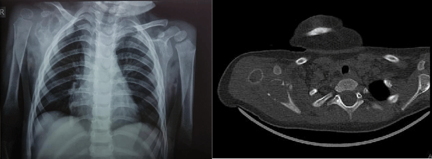
X-ray radiography showed a dislocation or arthritis in the right shoulder. Non-contrast Computed Tomographic (CT) scan of the right shoulder was performed which revealed the expansile lytic lesions along with cortex destruction and soft tissue extension of the scapula. However, the humorous bone had normal appearance (Figure 1). Right shoulder ultrasound confirmed the soft tissue component of the scapula cortex. At this stage, osteosarcoma, Ewing sarcoma, osteomyelitis and LCH, were considered as differential diagnoses.
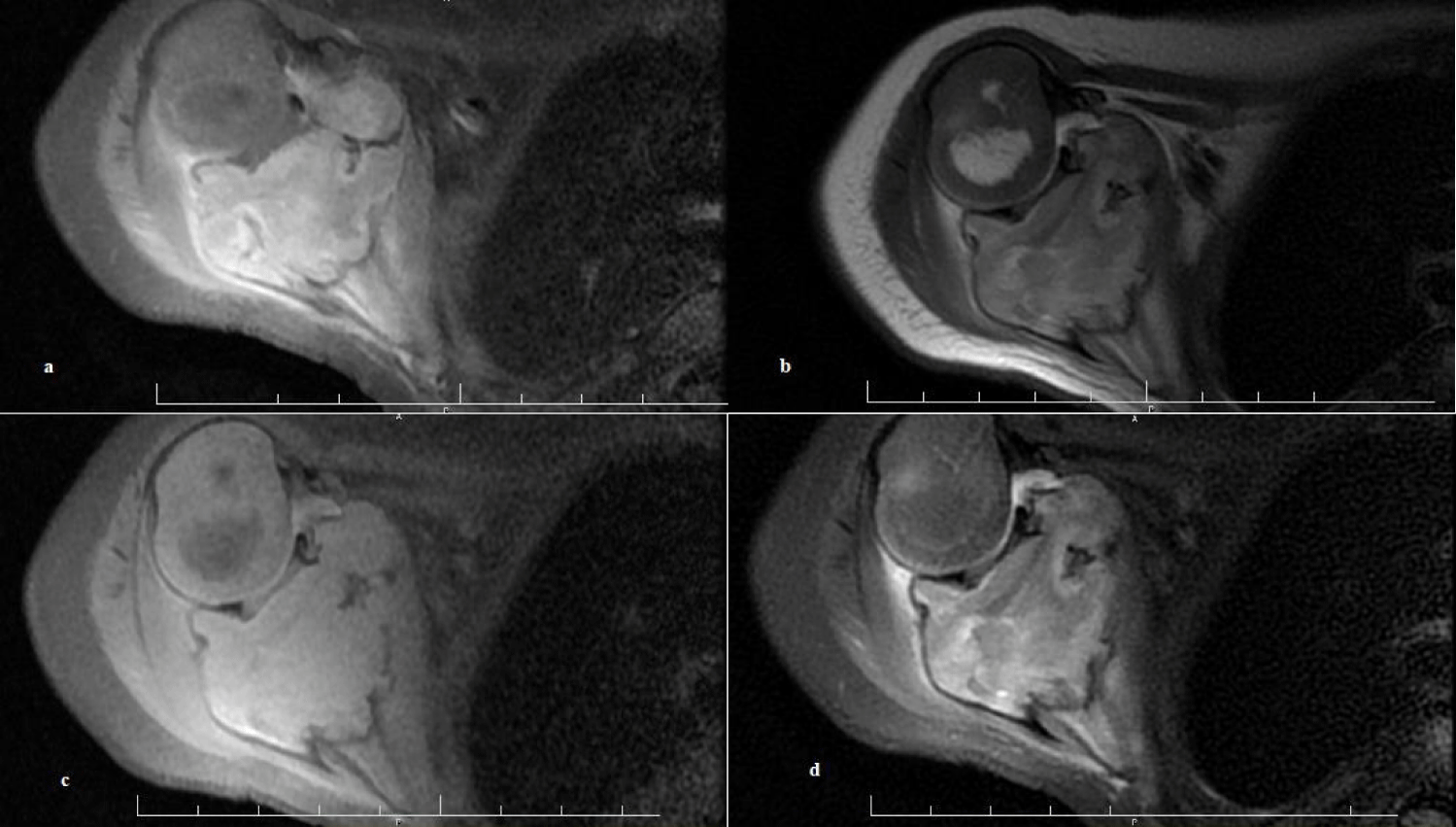
Right shoulder MRI with and/ or without contrast revealed an expansive hyper signal lesion in T2, iso signal to hyper signal intensity than muscle in T1, under the glenoid cavity in the body of scapula (3.4 cm axial and 5 cm craniocaudal diameters). After the contrast injection, a heterogeneous enhancement was observed. Various axillary lymph nodes were reported. The evidence expressed the possibility of a malignant tumor strongly (Figure 2).
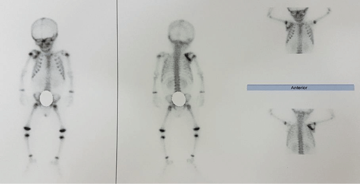
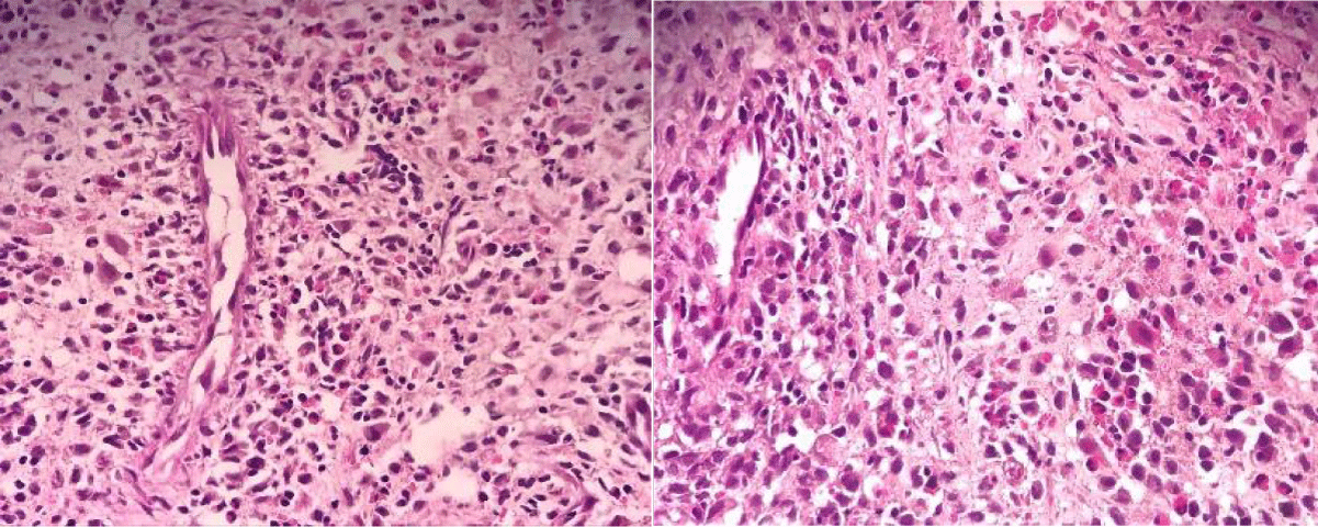
After IV injection of 7mCi of Tc-99m-MDP, the whole body bone scan showed an area of increased activity in the right scapula and confirmed the tumoral involvement. No other abnormal tracer uptake was noted in the rest of the skeleton. There was no evidence of skeletal metastasis (Figure 3). An incisional biopsy was taken from the mass of the scapula. The microscopic examination of the osteolytic lesion revealed a typical LCH. The tumor cells were strongly positive for Vimentin, CD68, S100, CD1a+, and 18% for Ki67, so immunohistochemistry findings confirmed LCH (Figure 4).
Laboratory findings demonstrated the hypochromic microcytic anemia and lymphocytosis. (WBC = 6700 (Neutrophil = 35.9, lymphocyte = 54.2), Hb = 12.3, Hct = 36.1, MCH = 26.5, MCHC = 34.1, MCV = 77.6, ESR = 83, CRP = 2+, and normal urine analysis). In addition, an abdominal sonography revealed no evidence of organ’s involvement.
Therefore, the patient was scheduled for chemotherapy (vinblastine 10 mg/m²/day IV over 1 minute q7 to 10 days) and prednisolone 30 mg daily together. After five months of follow-up, the size of the lesion and soft tissue edema was significantly decreased. No lymphadenopathy was detected at the MRI.
Discussion
LCH mostly occurs in girls under 10 years old. It presents as a uni to multifocal lesions. LCH affects multi organs or isolates in bone (especially skull), skin. The prevalence of LCH with the bone lesions; skull, jaw, long tubular bone, vertebrae, pelvis, and ribs were 51%, 30%, 17%, 13%, 13%, and 6%, respectively [9,10]. The unifocal, multifocal, and multisystemic types mostly present in adults, younger children, and infants, respectively [11]. The symptoms vary based on the site of involvement, mostly in the skin (with hearing loss, rash), CNS (with gate defect, ataxia, tremor, polyuria, polydipsia), and bone (with movement disabilities) [1,4].
This study presenting a rare case of granulomatosis disease, its manifestations, diagnostic and therapeutic approaches, in order to enhance the physicians’ knowledge to consider the LCH among the other differential diagnosis. In our study, LCH was presented as the bone lytic lesion at one of the rarest sites (scapula), disseminated to axillary lymph nodes, with a good outcome. This case was presented in an infant boy, which is also rarely common.
Among all bone tumors, scapula only accounts for 3%. Pandey, et al. [12] reported the unifocal LCH of the scapula in a 10-year-old boy, with pain and restriction of the right shoulder’s movement since one year previously. The biopsy reports (numerous histiocytes and pleomorphic eosinophils) and immunochemical findings (positive CD68 and S100) confirmed the diagnosis. The patient had good prognosis after the chemotherapy. Tsirakis, et al. [10] reported a multisystem LCH of the scapula and spleen in a young man that was diagnosed after radiological imaging, biopsies, and immunohistochemistry (S100, CD68, and CD1a). The patient received vinblastine, prednisolone, and 6-MP and had good prognosis.
Besides, a study presented a LCH of the clavicle in a 10-year-old girl in Iran, which was first misdiagnosed as osteomyelitis [13]. Similarly, Udaka, et al. [14] reported the LCH of the clavicle in a 25-year-old female. A 20 years study in Iran showed that only 20 cases (0.34% of all pathologic reports) were diagnosed as the LCH, which was more frequent in male adults [15].
Chemotherapy, chemo-immunotherapy, and radiotherapy play important roles in LCH treatment. In this regard, the medications such as vinblastine, methotrexate, 6-Mercaptopurine (6-MP), corticosteroids, and non-steroid anti-inflammatory drugs made a great revolution in treatment approaches, but the most effective therapeutic approach remains a medical challenge. Therefore, investigation of the clinical and paraclinical features of LCH can help to clarify its origin and possible associated factors to achieve the best diagnostic and therapeutic approaches [3,7,16].
References
- da Costa CE, Egeler RM, Hoogeboom M, Szuhai K, Forsyth RG, Niesters M, de Krijger RR, Tazi A, Hogendoorn PC, Annels NE. Differences in telomerase expression by the CD1a+ cells in Langerhans cell histiocytosis reflect the diverse clinical presentation of the disease. J Pathol. 2007 Jun;212(2):188-97. doi: 10.1002/path.2167. PMID: 17447723.
- Makras P, Polyzos SA, Anastasilakis AD, Terpos E, Papatheodorou A, Kaltsas GA. Is serum IL-17A a useful systemic biomarker in patients with Langerhans cell histiocytosis? Mol Ther. 2012 Jan;20(1):6-7. doi: 10.1038/mt.2011.239. PMID: 22215051; PMCID: PMC3255574.
- Rizzo FM, Cives M, Simone V, Silvestris F. New insights into the molecular pathogenesis of langerhans cell histiocytosis. Oncologist. 2014 Feb;19(2):151-63. doi: 10.1634/theoncologist.2013-0341. Epub 2014 Jan 16. PMID: 24436311; PMCID: PMC3926789.
- Nakamura T, Morimoto N, Goto F, Shioda Y, Hoshino H, Kubota M, Taiji H. Langerhans cell histiocytosis with disequilibrium. Auris Nasus Larynx. 2012 Dec;39(6):627-30. doi: 10.1016/j.anl.2012.01.003. Epub 2012 Feb 10. PMID: 22326120.
- Babaniamansour S, Aliniagerdroudbari E, Niroomand M. Glycemic control and associated factors among Iranian population with type 2 diabetes mellitus: a cross-sectional study. J Diabetes Metab Disord. 2020 Jul 8;19(2):933-940. doi: 10.1007/s40200-020-00583-4. PMID: 33520813; PMCID: PMC7843766.
- Gabbay LB, Leite Cda C, Andriola RS, Pinho Pda C, Lucato LT. Histiocytosis: a review focusing on neuroimaging findings. Arq Neuropsiquiatr. 2014 Jul;72(7):548-58. doi: 10.1590/0004-282x20140063. PMID: 25054989.
- Van’t Hooft I, Gavhed D, Laurencikas E, Henter JI. Neuropsychological sequelae in patients with neurodegenerative Langerhans cell histiocytosis. Pediatr Blood Cancer. 2008 Nov;51(5):669-74. doi: 10.1002/pbc.21656. PMID: 18623210.
- Babaniamansour P, Mohammadi M, Babaniamansour S, Aliniagerdroudbari E. The Relation between Atherosclerosis Plaque Composition and Plaque Rupture. J Med Signals Sens. 2020 Nov 11;10(4):267-273. doi: 10.4103/jmss.JMSS_48_19. PMID: 33575199; PMCID: PMC7866947.
- Aricò M, Nichols K, Whitlock JA, Arceci R, Haupt R, Mittler U, Kühne T, Lombardi A, Ishii E, Egeler RM, Danesino C. Familial clustering of Langerhans cell histiocytosis. Br J Haematol. 1999 Dec;107(4):883-8. doi: 10.1046/j.1365-2141.1999.01777.x. PMID: 10606898.
- Tsirakis G, Kaparou M, Kanellou P, Kontakis G, Alexandrakis M. Langerhans cell histiocytosis in the right scapula in a young man. International Journal of Case Reports and Images. 2012;3:16. https://bit.ly/2PNkUXh
- Yoon JH, Park HJ, Park SY, Park BK. Langerhans cell histiocytosis in non-twin siblings. Pediatr Int. 2013 Jun;55(3):e73-6. doi: 10.1111/ped.12034. PMID: 23782385.
- Pandey R, Bhayana H, Rajnish R, Dhammi I, Jain A, Haq R-U. Langerhans cell histiocytosis of the scapula-diagnosis & treatment options. Coluna/Columna. 2017;16:240-3. https://bit.ly/3a426d6
- Sayyahfar S, Ansari S, Rahbar M, Zarei E. Langerhans cell histiocytosis of the clavicle in a 10-years-old girl. Iranian journal of Pediatric Hematology and Oncology. 2017;7(4):260-263. https://bit.ly/3mEE2Cu
- Udaka T, Susa M, Kikuta K, Nishimoto K, Horiuchi K, Sasaki A, Kameyama K, Nakamura M, Matsumoto M, Chiba K, Morioka H. Langerhans Cell Histiocytosis of the Clavicle in an Adult: A Case Report and Review of the Literature. Case Rep Oncol. 2015 Oct 16;8(3):426-31. doi: 10.1159/000441415. PMID: 26600774; PMCID: PMC4649755.
- Atarbashi Moghadam S, Lotfi A, Piroozhashemi B, Mokhtari S. A Retrospective Analysis of Oral Langerhans Cell Histiocytosis in an Iranian Population: a 20-year Evaluation. J Dent (Shiraz). 2015 Sep;16(3 Suppl):274-277.. PMID: 26535408; PMCID: PMC4623830.
- Gavhed D, Laurencikas E, Akefeldt SO, Henter JI. Fifteen years of treatment with intravenous immunoglobulin in central nervous system Langerhans cell histiocytosis. Acta Paediatr. 2011 Jul;100(7):e36-9. doi: 10.1111/j.1651-2227.2010.02125.x. Epub 2011 Jan 11. PMID: 21166862.
Content Alerts
SignUp to our
Content alerts.
 This work is licensed under a Creative Commons Attribution 4.0 International License.


