> Medicine Group. 2021 June 03;2(6):443-446. doi: 10.37871/jbres1257.
Extra Articular False Ankylosis Following Fusion of Mandible to Zygomatic Bone and Maxilla Due to Ossifying Fibroma-A Rare Case Report
Abhay Nilkanth Datarkar1*, Hema Anukula1, Suraj Parmar1, Anshul Rai2 and Suchitra Gosavi3
2Associate Professor Department of Dentistry, All India Institute of Medical Sciences, Bhopal, India
3Professor and Head Department of Oral and Maxillofacial Pathology Government Dental College and Hospital, Nagpur, India
- Ossifying fibroma
- False ankylosis
- Extra Articular Ankylosis (EAA)
- Trismus
- Mandibular Zygomatic Fusion(MZF)
Abstract
Trismus is a disturbing condition with varied etiology, one of them is Extra articular ankylosis. The extra articular ankylosis has been reported in the literature as a fusion of the coronoid process to the zygomatic bone or to the skull base. However, there are a very few cases reported in the literature that highlights a pathologic tumor of mandible leading to extra-articular ankylosis and subsequently resulting in trismus. We hereby present a unique case of young adult having restricted mouth opening due to the bony fusion of the mandibular ramus to the zygomatic bone and zygomaticomaxillary buttress region due to a pathologic tumor diagnosed as Ossifying Fibroma.
Introduction
Trismus is a disturbing and debilitating condition for the patients as it affects the function of mastication that subsequently results in malnutrition. Amongst the numerous causes of trismus, the one presented in this manuscript is Ankylosis, specifically the Extra articular variant. The extra articular or the False Ankylosis has been reported in the literature as a fusion of the coronoid process to the zygomatic bone or to the skull base [1].
The most common etiology of the False ankylosis reported is due to trauma causing fracture of the Zygomatico Maxillary Complex (ZMC) and coronoid process which were undiagnosed and untreated [2]. However, there are a very few cases reported in the literature that highlights a pathologic tumor of mandible posing an etiological factor for extra-rticular ankylosis. Those reported are mainly the tumors of coronoid process [3] leading to trismus. There is hardly any case of bony tumor of Ramus and Angle region of mandible reported which results in reduction of mouth opening due to fusion with the zygomatic bone.
We hereby present a rare case entity of young adult having restricted mouth opening due to the bony fusion of the mandibular ramus to the zygomatic bone and zygomatico buttress region due to a pathologic tumor. This kind of condition is unique and hardly reported.
Case Report
A 14-year-old male patient reported with chief complaint of reduction in mouth opening since four and half years. Patient’s initial mouth opening was approximately equal to his 4 fingers width. Patient noticed sudden onset of swelling over his left side of face and decrease in the mouth opening for which he visited a private hospital where he was operated for the release of fusion of ramus to zygomatic arch through an intraoral approach achieving 40mm mouth opening three years back and he was suggested a regular physiotherapy. Patient now reported to us with nil mouth opening leading to recurrence of condition. Patient gives no history of trauma or habits like betel nut or tobacco chewing.
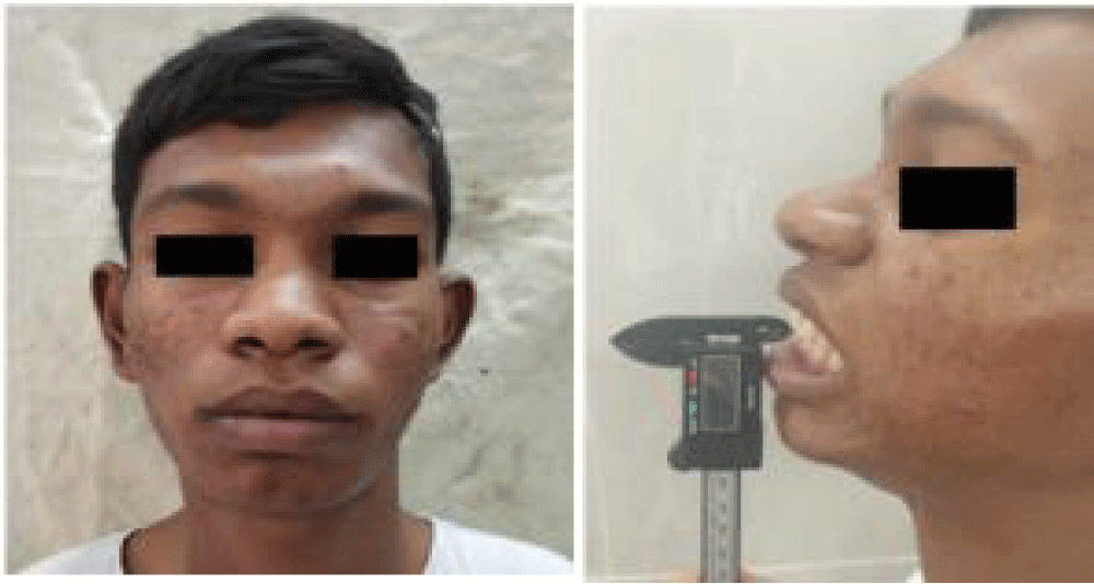
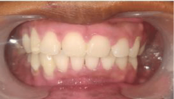
Clinical examination revealed apparently symmetrical face as shown in (Figure 1). Mouth opening was nil (Figure 2) with mild protrusive and lateral excursion movements. On palpation soft tissue overlying left malar region was firm. There was no tenderness over left malar region. Intraoral examination revealed that occlusion was not deranged and there was no abnormality detected in intraoral soft tissues (Figure 3).
Investigations
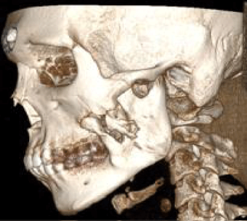
Panoramic radiograph revealed the radio opacity that was bridging from anterior border of ramus to zygomatic bone. Bilateral condyle appeared normal. Computed tomography scan revealed the hyperdense area noticed from anterior border of ramus to zygomatic buttress region forming bridge like structure (Figure 4). Normal joint space and normal condylar shape was appreciated. After biopsy, a treatment plan of excision of the bony tumor was planned under general anesthesia.
The clinical differential diagnosis are as follows;
• Osteochondroma/ chondroma of coronoid
• Osteoma of coronoid
• Fibro osseous lesion
• Coronoid – Pterygoid fusion
• Zygomatico coronoid fusion
Treatment
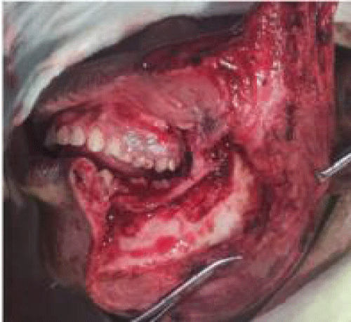
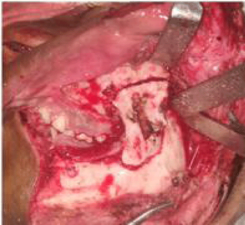
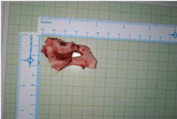
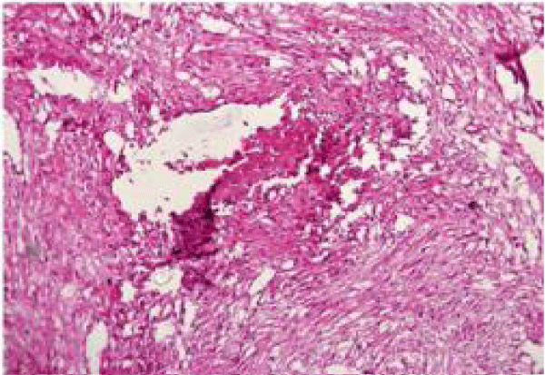
After obtaining the informed risk consent the patient was subjected to surgery and awake fiberoptic bronchoscope guided intubation was performed. Robson incision was marked over left side. The incision was placed through the skin and subcutaneous tissue. Layer-wise dissection was carried out through platysma to superficial layer of deep cervical fascia. Facial artery was identified and ligated. The periosteum over the lower border of mandible was dissected and subperiosteal dissection was carried out. The zygomatic buttress to posterior border of ramus was exposed (Figure 5). Osteotomy was marked with safe margin of 1 cm surrounding the pathology and excision of pathology was done (Figure 6). Ipsilateral coronoidectomy was done. The excised specimen (Figure 7) was sent for histopathological examination which showed mostly mature bone with cellular fibroblastic stroma with vague storiform pattern with foci of bone formation, suggestive of ossifying fibroma.
The mouth opening after excision of pathology and ipsilateral coronoidectomy was 32 mm. Contralateral coronoidectomy was also done intraorally and 45 mm of mouth opening was achieved. Drain was secured and layer wise suturing was done with 3-0 vicryl and 4-0 prolene extra orally and intraorally suturing was done using 3-0 vicryl. The patient was given Inj Amoxicillin and Clavulanic Acid 1.2 gm IV BD, Inj. Metronidazole 500 mg IV TDS and Inj. Diclofenac Sodium 25mg IM BD during the postoperative period.
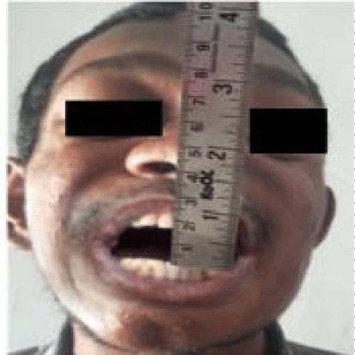
The active mouth opening exercises were started from third post-operative day and patient was discharged on the fifth post-operative day five. Patient is on regular follow-up since 1 year and at present patient mouth opening was 18 mm between incisal edge of upper and lower central incisor (Figure 8) and there is no sign of recurrence. Patient is encouraged to do regular mouth opening exercises. The histopathological diagnosis of excised specimen was Ossifying Fibroma.
Discussion
Amongst the various causes of trismus, one is fusion of mandible to maxilla, whether it is Temporomandibular Joint ankylosis or ankylosis of coronoid process to Zygomatic bone [4]. However, fusion of Mandibular Body-Ramus to the Zygomatic bone is very rare. The main causes of extra-articular ankylosis such as fusion of the coronoid process to the zygomatic bone [5], bone graft migration [6], abnormal coronoid elongation due to chondroma, osteochondroma, Osteoma or due to exostosis is commonly reported in the literature [7]. Miyamoto S, et al. [8] reported that the causes of extra-articular ankylosis are infection, trauma, post radiation therapy or space occupying lesions. Some authors believe that the osteogenic potential is due to surgery induced metaplastic changes in the connective tissue component [9]. However, the ossifying fibroma as an etiologic factor for false ankylosis has not been documented in the literature.
A painless limitation in mouth opening is common in patients with extra-articular ankylosis. Generally, these patients do not have limitation of protrusive and lateral jaw movements. Proper clinical and Radiographic Diagnosis is very much important.
Hence, obtaining a CT scan helps in arriving at a provisional diagnosis and formulating the treatment plan due to the detailed anatomic information that it provides.
OF is a benign fibro-osseous lesion that occurs in children, adolescents and adults [10]. The mandible is most commonly affected by OF [11], but extension of the lesion from the mandible to the zygoma creating restriction in mouth opening is rare.False ankylosis due to ossifying fibroma is the rarest. It can be considered as a differential diagnosis for long standing Trismus without history of trauma or submucous fibrosis Various treatment modalities like application of brisement force under general anesthesia, coronoidectomy, adjuvant radiation therapy limiting the recurrent heterotopic bone after surgical excision have been reported. Excision of tumor and active physiotherapy, however is the modality of choice [12]. Literature suggests that Ossifying fibroma requires radical surgery because of the tendency for recurrence and possibility of malignant transformation [13]. In the present case, excision was successfully done followed by bilateral coronoidectomy to achieve adequate mouth opening with adjuvant post-operative physiotherapy.
Considering the previous history of intra oral release of the lesion leading to recurrence at a 3 year period a radical extra oral approach by using Robson incision was taken. This approach provided good exposure of the surgical site and helps in easy surgical excision; however, it is associated with the disadvantage of poor aesthetics in the form of extra oral scar to the young boy but can be minimized by putting the incision under the lower border of mandible. Finally early surgical intervention, with proper Antibiotics and Fluid therapy is required for good result.
Regular post operative physiotherapy and a long term Follow up of the patient is very much important.
Conclusion
We presented a unique case of trismus due to fusion of zygomatic buttress to the ramus and body of mandible.This patient had a history of recurrence after previous surgery. Radical approach for the complete excision of such tumour along with regular physiotherapy is the key for prevention of recurrence. The histopathological diagnosis of bony tumour of ossifying fibroma leading to trismus and inability to open mouth is very unique presentation in this case report.
References
- Karakasis D, Triantafyllidou E, Kavadia S. Extra-articular ankylosis of the coronoid processes to the base of the skull. A case report. J Craniomaxillofac Surg. 1989 Jan;17(1):46-9. doi: 10.1016/s1010-5182(89)80127-1. PMID: 2915046.
- Tippu SR, Rahman F. Heterotopic calcification: A cause for zygomatico- coronoid ankylosis. Biomed Res. 2011;22.
- Lan T, Liu X, Liang PS, Tao Q. Osteochondroma of the coronoid process: A case report and review of the literature. Oncol Lett. 2019 Sep;18(3):2270-2277. doi: 10.3892/ol.2019.10537. Epub 2019 Jun 27. PMID: 31452728; PMCID: PMC6676659.
- Lee SM, Baek JA, Kim Y. Ankylosis of the Coronoid Process to the Zygomatic Bone: A Case Report and Review of the Literature. J Oral Maxillofac Surg. 2019 Jun;77(6):1230.e1-1230.e11. doi: 10.1016/j.joms.2018.10.006. Epub 2018 Oct 18. PMID: 30439329.
- Vanhove F, Dom M. Zygomatico-coronoid ankylosis: a case report. Int J Oral Maxillofac Surg. 1999 Aug;28(4):258-9. PMID: 10416891.
- Emekli U, Arslan A, Onel D, Kesim SN. Extra-articular ankylosis of the mandible caused by possible migration of bone grafts. Ann Plast Surg. 2003 Oct;51(4):435-6. doi: 10.1097/01.SAP.0000091209.05975.49. PMID: 14520077.
- Iqbal S, Hamid AL, Purmal K. Unilateral coronoid hyperplasia following trauma: a case report. Dent Traumatol. 2009 Dec;25(6):626-30. doi: 10.1111/j.1600-9657.2009.00830.x. Epub 2009 Oct 14. PMID: 19843134.
- Miyamoto S, Takushima A, Momosawa A, Ozaki M, Harii K. Transzygomatic coronoidectomy as a treatment for pseudoankylosis of the mandible after transtemporal surgery. Scand J Plast Reconstr Surg Hand Surg. 2008;42(5):267-70. doi: 10.1080/02844310701270265. PMID: 18788051.
- Kallalli BN, Rawson K, Manugutti A, Swapna S. Right zygomatico coronoid ankylosis: A rare clinical entity. J Indian Acad Oral MedRadiol 2014;26:323-226. doi: 10.4103/0972-1363.145019
- Akcam T, Altug HA, Karakoc O, Sencimen M, Ozkan A, Bayar GR, Gunhan O. Synchronous ossifying fibromas of the jaws: a review. Oral Surg Oral Med Oral Pathol Oral Radiol. 2012 Nov;114(5 Suppl):S120-5. doi: 10.1016/j.oooo.2011.08.007. Epub 2012 Feb 25. PMID: 23063387.
- Ojo MA, Omoregie OF, Altini M, Coleman H. A clinico-pathologic review of 56 cases of ossifying fibroma of the jaws with emphasis on the histomorphologic variations. Niger J Clin Pract. 2014 Sep-Oct;17(5):619-23. doi: 10.4103/1119-3077.141429. PMID: 25244274.
- Triantafillidou K, Venetis G, Karakinaris G, Iordanidis F. Ossifying fibroma of the jaws: a clinical study of 14 cases and review of the literature. Oral Surg Oral Med Oral Pathol Oral Radiol. 2012 Aug;114(2):193-9. doi: 10.1016/j.tripleo.2011.07.033. Epub 2012 Feb 25. PMID: 22776732.
- Liu Y, Wang H, You M, Yang Z, Miao J, Shimizutani K, Koseki T. Ossifying fibromas of the jaw bone: 20 cases. Dentomaxillofac Radiol. 2010 Jan;39(1):57-63. doi: 10.1259/dmfr/96330046. PMID: 20089746; PMCID: PMC3520406.
Content Alerts
SignUp to our
Content alerts.
 This work is licensed under a Creative Commons Attribution 4.0 International License.


