Medicine Group . 2022 February 14;3(2):170-173. doi: 10.37871/jbres1415.
Discussion of MRI Findings with Surgical Correlation in Vaginal and Cervical Agenesis: A Short Review of Mullerian Duct Anomalies
Kritika Sharma, Rajaram Sharma*, Tapendra Nath Tiwari and Saurabh Goyal
Abstract
Magnetic Resonance Imaging (MRI) is the modality of choice and is considered a highly accurate tool for the evaluation of Müllerian duct anomalies to characterize the uterine and vaginal anomalies [1,2]. In clinical practice, the MRI examination is used by the clinician to reach the diagnosis, for assessing the abnormalities and planning for surgery or future intervention. The information provided by other examinations like pelvic examination, sonography, hysterosalpingography can be confirmed and complemented by MRI. The case described here illustrates the standard MRI findings of vaginal and cervical agenesis with clinical and surgical correlation and revisits the uses of MRI.
Introduction
Congenital uterine malformations are due to defective fusion of Mullerian ducts in the developing embryo. These anomalies are in isolation or in combination with other urological malformations. The prevalence of female congenital malformations in the general population is up to 7%. Females with these kinds of abnormalities usually present during menarche age, when there is an absence of onset of menses, cyclical abdominal pain and depending upon the severity, the presentation could be late during the fertility period. Cervical agenesis is a rare Mullerian anomaly with an incidence of 1 in 80,000 females. It represents 3% of all uterine anomalies. It is very often associated with vaginal atresia (< 50%). A functional uterus with cervical and vaginal atresia is very rare; less than 20 cases are reported yet in literature [3]. It is important to classify these anomalies for easy diagnosis and appropriate preoperative assessment, and for that, MRI is the best modality.
Case Presentation
A 15-years old female came to our gynaecology department with a history of primary amenorrhea and cyclic lower abdominal pain from the last year. She had normal vital parameters; however, their tenderness could be elicited in the lower abdomen. On physical examination, there were normal secondary sexual characteristics with appropriate growth. In external genitalia labia majora, labia minora were normal, but no vaginal opening was present.
Routine blood and serological investigations were unremarkable, including normal serum Follicle-Stimulating Hormone (FSH), Luteinizing Hormone (LH), and prolactin levels.
Investigation
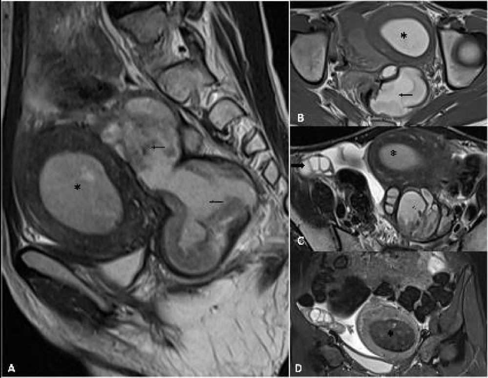
Trans-abdominal ultrasound demonstrated a deformed uterus with endometrial collection having low-level internal echoes with non-visualization of the cervix and vaginal canal. A large tubular cystic lesion with internal echoes and incomplete septations was observed in the left adnexa representing the hydrosalpinx. The left ovary was not be visualized separately from this lesion. Based on the features mentioned above, a diagnosis of tubo-ovarian mass-made. During the evaluation of tumour markers, the CA-125 was raised (400 U/ml). An MRI scan of the pelvis was performed for further evaluation, which revealed a bulky uterus (7.1 x 5.2 x 7.0 cm) containing haemorrhagic endometrial collection (35 cc). Cervix and vagina could not be identified on MRI. A large, well defined, lobulated lesion (4.7 x 4.5 x 9.0 cm) was noted in the left adnexal region, which appeared hyperintense on the T1W image, iso to hyperintense on T2W/STIR images. The lesion also had internal organized blood products and septations. The left fallopian tube was grossly dilated and filled with blood products (7 mm in diameter). The haemorrhagic collection is also seen in the pouch of Douglas, and the right ovary is ectopically visualized in the right iliac fossa (Figure 1A-D) (Video 1). A diagnosis of left hematosalpinx with haemorrhagic tubo-ovarian mass was made.
Differential Diagnosis
Other differentials like neoplastic ovarian lesion and pelvic tuberculosis were also considered in view of raised CA-125 levels and adhered fallopian tube and ovary, respectively, in the pre-surgical meet. However, considering the age of the patient, physical examination findings and imaging features, these were considered as less likely diagnoses.
Treatment
After taking the informed written consent and explaining the possibility of hysterectomy to the parents, the patient underwent an exploratory laparotomy. Intraoperatively, there was a left-sided hematosalpinx and hematometra in a blind-ended, globular shaped, thick-walled cystic structure with internal hyperintense collection with no obvious outline of cervix and vagina suggestive of the uterine body (Figure 2A-C). There was agenesis of the cervix and vagina. The left ovary was adhered to the haematosalpinx and had to be removed. On the right side, the fallopian tube was streak like and hypoplastic with the right ectopic ovary. Removal of the uterine body and left salpingectomy were performed.
Outcome and Follow-up
After the uneventful postoperative period, the patient was discharged and kept on symptomatic medications. Vaginoplasty is has been planned for the near future, as the patient is not giving consent for the same at present.
Discussion
The female reproductive system, which consists of external genitalia (originated from ectoderm), gonads (from mesoderm origin) and the duct system between these two, are also mesoderms in origin. The paired Mullerian duct system develops to form fallopian tubes, the uterine corpus, cervix, and the upper third of the vagina. The lower two-thirds of the vagina is derived from the urogenital sinus of the ectoderm, which eventually fuses with the upper third. Any abnormality in this process leads to Mullerian duct anomalies. These anomalies are classified into four groups, a) absence of any part, i.e. agenesis, b) problem in lateral fusion, c) problem in vertical fusion, d) any combination of the above. The classification of vaginal agenesis is as follows, a) partial agenesis with midline functional uterus and cervix, b) complete agenesis of the vagina with a functional uterus but no cervix, c) complete vaginal agenesis with rudimentary uterine segment and functioning endometrium (our patient falls in this category). Cervical atresia can be observed in three configurations: 1) total absence of the cervix; 2) a cervix containing only stromal tissue with no cervical canal; or 3) a cervix without canalization but with small inclusion. Any abdominal or pelvic pain and history of primary amenorrhea in a pubescent girl must evoke the suspicion of possible obstructive genital anomaly. Clinically it may present with obstructive symptoms, cyclical lower abdominal pain, or maybe asymptomatic depending on the functional status of the endometrium [4]. The clinical examination helps identify lower genital tract anomalies like imperforate hymen or the presence of external genitalia or vaginal opening. A large number of women with these anomalies have associated anomalies of the urinary tract and skeleton, but most of them were found to have standard chromosomal patterns and normal ovaries. This classification helps in making surgical decisions as the patient lies in the first category of vaginal agenesis in which only external opening was needed to be created for menstrual discharge rest of the other categories require hysterectomy with or without vaginoplasty. While it is impossible to create a cervix with the functional endocervical canal to allow pregnancy through the vaginal canal can be made.
Reconstructive surgery includes uterovaginal anastomosis, which is the preferred treatment for cervical agenesis or atresia, cervical obstruction or a fibrous cord. The aim of these reconstructive surgeries is to provide a passage for menstruation to relieve pain and try to preserve reproductive potential. But the patient with cervical agenesis or cervical fragmentation are usually poor candidates for canalization; for them, hysterectomy is the treatment of choice [5].
After all the controversies about the treatment procedures, it is being concluded that preoperative evaluation is essential to provide relevant information about the remnant cervix and vagina so that all the risks and benefits of the possible procedure can be evaluated. Hysterectomy was the eventual treatment for cervical agenesis because of the higher complications of recanalization of the cervix and the unlikelihood of a viable pregnancy [6]. However, hysterectomy might be necessary when the conservative treatment fails. Therefore, for this evaluation, MRI has the best potential for both functional and morphological characteristics. Sagittal images allow a clear assessment of the appearance of the corpus uteri, cervix, and vagina, as their long axes are usually on a single plane. Further uterine walls are also well-differentiated on MRI into three distinct zones with a low-intensity band. Blood or blood products are considered as a definite sign of functional endometrium that can also be observed on MRI. Short mature cervix, cervical lip and endocervical canal can be easily demonstrated on MRI. The studies of Reinhold, et al. [7] showed excellent agreement between MRI and surgical findings in patients with MRKH syndrome, cases of cervical agenesis or dysgenesis. Recent reports have also suggested that MRI offers the best support in achieving a preoperative diagnosis.
Learning Points
- Cervical and vaginal agenesis with non communicating rudimentary horn is a very rare entity which is often diagnosed late in the disease course.
- MRI is a very useful tool in the evaluation of patients with suspected Mullerian duct anomalies as it offers sufficient information for surgical management.
- CA-125 level may be raised in non-neoplastic hemorrhagic tubo-ovarian lesions.
References
- Troiano RN, McCarthy SM. Mullerian duct anomalies: imaging and clinical issues. Radiology. 2004 Oct;233(1):19-34. doi: 10.1148/radiol.2331020777. Epub 2004 Aug 18. PMID: 15317956.
- Carrington BM, Hricak H, Nuruddin RN, Secaf E, Laros RK Jr, Hill EC. Müllerian duct anomalies: MR imaging evaluation. Radiology. 1990 Sep;176(3):715-20. doi: 10.1148/radiology.176.3.2202012. PMID: 2202012.
- Jacob JH, Griffin WT. Surgical reconstruction of the congenitally atretic cervix: two cases. Obstet Gynecol Surv. 1989 Jul;44(7):556-69. doi: 10.1097/00006254-198907000-00011. PMID: 2662083.
- Deffarges JV, Haddad B, Musset R, Paniel BJ. Utero-vaginal anastomosis in women with uterine cervix atresia: long-term follow-up and reproductive performance. A study of 18 cases. Hum Reprod. 2001 Aug;16(8):1722-5. doi: 10.1093/humrep/16.8.1722. PMID: 11473972.
- Acién P, Acién M, Sánchez-Ferrer M. Complex malformations of the female genital tract. New types and revision of classification. Hum Reprod. 2004 Oct;19(10):2377-84. doi: 10.1093/humrep/deh423. Epub 2004 Aug 27. PMID: 15333604.
- Al-Jaroudi D, Saleh A, Al-Obaid S, Agdi M, Salih A, Khan F. Pregnancy with cervical dysgenesis. Fertil Steril. 2011 Dec;96(6):1355-6. doi: 10.1016/j.fertnstert.2011.09.029. Epub 2011 Oct 7. PMID: 21982728.
- Reinhold C, Hricak H, Forstner R, Ascher SM, Bret PM, Meyer WR, Semelka RC. Primary amenorrhea: evaluation with MR imaging. Radiology. 1997 May;203(2):383-90. doi: 10.1148/radiology.203.2.9114092. PMID: 9114092.
Content Alerts
SignUp to our
Content alerts.
 This work is licensed under a Creative Commons Attribution 4.0 International License.


