Medicine Group . 2022 March 08;3(3):221-226. doi: 10.37871/jbres1426.
Lead Extraction and Re-Extractions - Inherent Parts of Permanent Pacing in Children and Young Adults
Paweł Stefanczyk1, Anna Polewczyk2,3, Dorota Nowosielecka1*, Lukasz Tulecki4, Maria Miszczak-Knecht5, Wojciech Jachec6, Andrzej Kleinrok1,7, Katarzyna Bieganowska5 and Andrzej Kutarski8
2Faculty of Medicine and Health Sciences, The Jan Kochanowski University, Kielce, Poland
3Department of Cardiac Surgery, Świętokrzyskie Cardiology Center, Kielce, Poland
4Department of Cardiac Surgery, The Pope John Paul II Province Hospital of Zamość, Poland
5Department of Cardiology, The Children’s Memorial Health Institute, Warsaw, Poland
6Silesian Medical University, 2nd Department of Cardiology, Zabrze, Poland
7Medical College, Department of Physiotherapy, University of Information Technology and Management, Rzeszow, Poland
8Department of Cardiology, Medical University, Lublin, Poland
Abstract
Children require often replacement of leads even several times. Repeated extraction in this group nay be challenging. We report the case of a 22-year-old man with the first pacemaker implanted in the first year of life, after Transvenous Lead Extraction procedure (TLE) and implantation of a dual-chamber pacemaker in the age of 12 years, who was admitted to the reference center for repeated TLE and to replace the entire pacing system. The presence of complete venous occlusion, lead strain and strong lead-to-lead adherence with calcified connecting tissue scar effected TLE complicity and needed utility of numerous tools and atypical technique and tricks among of them simultaneous extraction of strongly connected each one leads together via one tool showed to be crucial.
Background
Permanent pacing in children is a big challenge because they often require replacement of leads even several times [1-8]. Growth of the body causes lead length deficiency, least strain and continuous pooling of lead which effected lead failures due to potential lead dislodgment and break of lead isolation [8-13]. Moreover, high physical activity might contribute increased risk of conductor break [8-13]. Additional lead implantation is accepted option but sometimes impossible due to venous occlusion [14,15] and may to generate future negative consequences [16-20].
Therefore, children and juveniles with CIED system requires in most of cases even several replacements during their lifetime and Transvenous Lead Extraction (TLE) is inherent part of electrotherapy in them [9,21-26]. Such procedures in this group of patients are more difficult due to intensive connecting tissue scar reaction for lead presence and earlier its calcification [21-26]. Experience with difficult extraction and repeated lead extraction in adults were describe [27,28] but in children and juveniles experience is limited.
We report the case extremely difficult lead re-extraction in 22-year-old man with 21-y-long pacing history and previous lead replacement. Both procedures were caused with bead dysfunction caused by body growth.
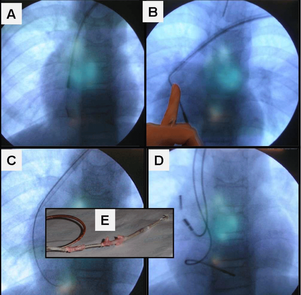
History single chamber pacemaker was implanted in 1997 due to a third-degree atrioventricular block. The next procedure in 2008 at the age of 12 due to lead dysfunction (Figure 1) where the longstanding (11-y old) lead was extracted under general anesthesia with mechanical telescoping sheaths (Byrd dilators) and a new Dual-Chamber Pacemaker (DDD) was implanted.
Due to the dislocation of the atrial electrode, the patient required the electrode replacement a few days after the last procedure. Then, a slight swelling of the left upper limb was also observed immediately after the procedure. For that reason, the Doppler ultrasound examination was performed, which showed slow blood flow through the subclavian vein, without thrombus. In the consequence, anticoagulant prophylaxis - low molecular weight heparin was applied which improved the problem.
The second, described procedure was performed in a hybrid operating room, under general anesthesia, with cardiosurgical team and transesophageal echocardiography monitoring. As the patient was pacemaker-dependent, temporary pacing via femoral access was used. Radiography of the chest before the procedure revealed two leads – one lead in the right ventricle, tightly taut and pulling the tip of the right ventricle, one atrial lead - seemingly loose and complete occlusion of the left axillary and subclavian vein after previous extraction. The first problem appeared at the very beginning. In order to stabilize both leads during the dissection, locking stylets were applied.
The first one step (conventional). The first technique for lead extraction was using non-powered mechanical systems with polypropylene telescoping dilators (Byrd Dilator Sheaths, Cook Medical Inc., USA). By using dilators for each of the electrodes separately, it was possible to move to the area of the beginning of the superior vena cava, where the presence of a strong lead-to-lead adherence disabled to continue dilatation.
The second step (conventional). The similar attempt was done with the next one lead with the same effect.
Third step. In the next stage, the first atypical attempt was made to separate the leads by means of two sheath simultaneously, the operation which can be compared to scissors work, but this also did not bring the expected effect. This method has already been tried and described earlier [29].
Fourth step. The last trick with polypropylene telescoping dilators was the use of the largest diameter sheath (orange outer) and an attempt to remove both electrodes simultaneously to dissect them from the vein wall, but due to massive adhesion and calcification, it also proved to be ineffective (Figure 1).
Fifth step. When the polypropylene telescoping sheaths were ineffective, a powered mechanical sheath system (Evolution Mechanical Dilator Sheath, Cook Medical Inc., USA) was used as a second line tool in our center. One of the lead was covered with green sheath and Evolution over the second one. It was petty progress but there was winding of the non-extracted lead conductor over Evolution sheath, which resulted in the destruction of the leads.
Sixth stem. In the next step, the leads and tools were changed (replaced), but that also did not work and the leads were not separated from each other.
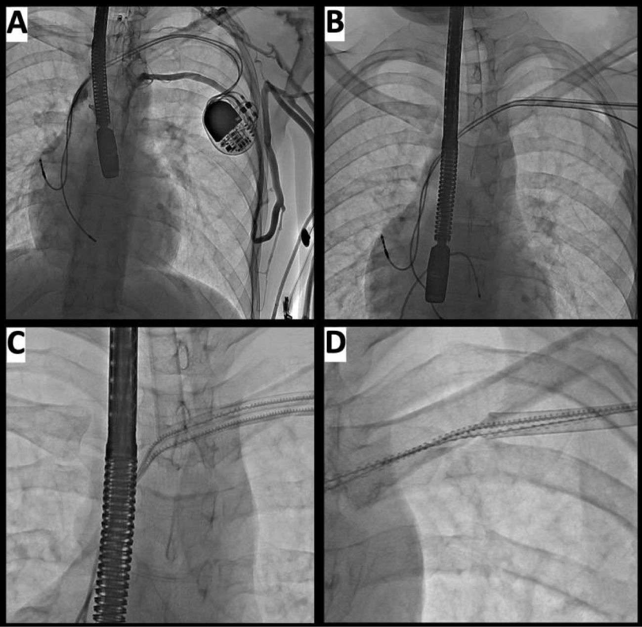
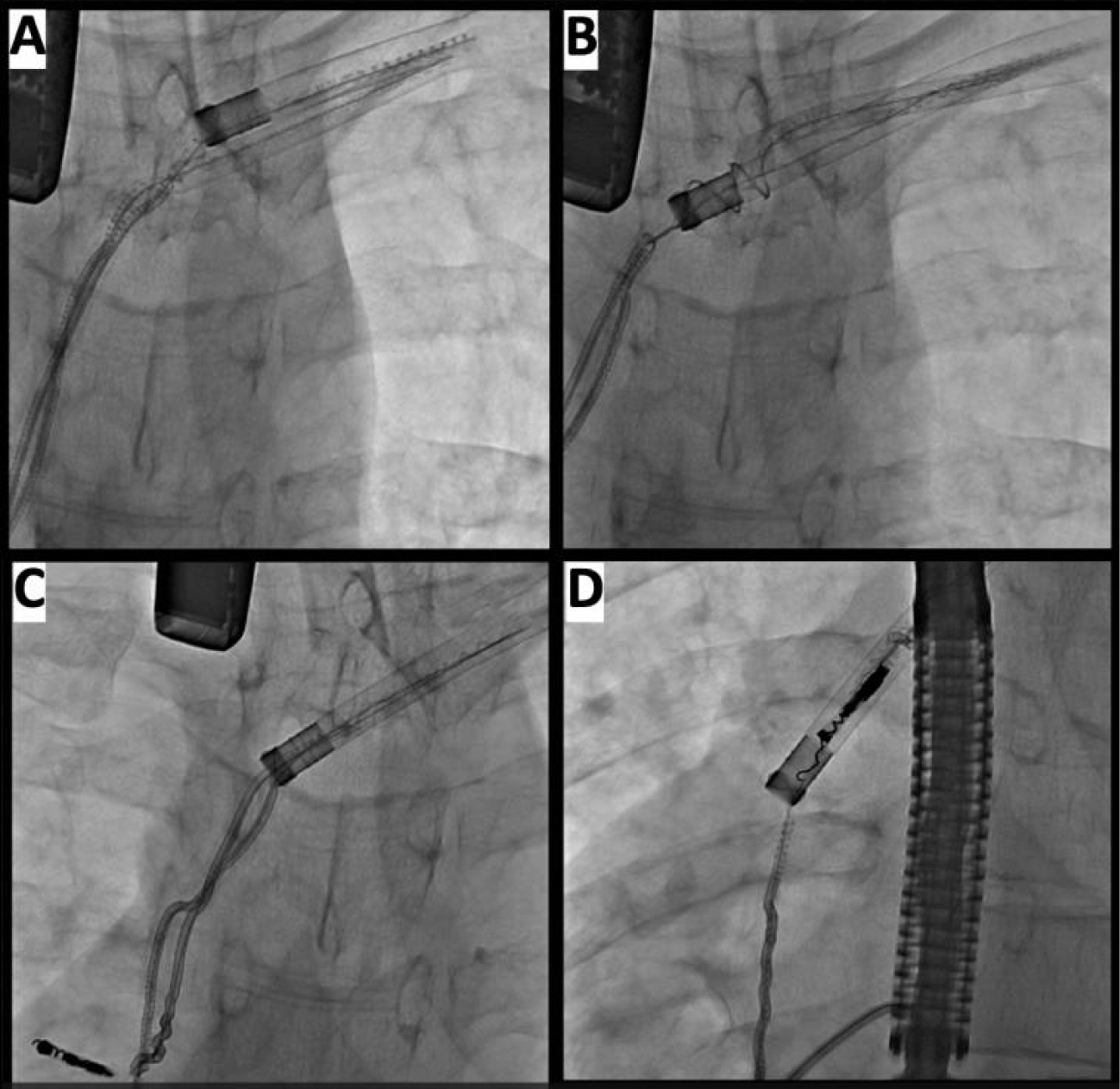
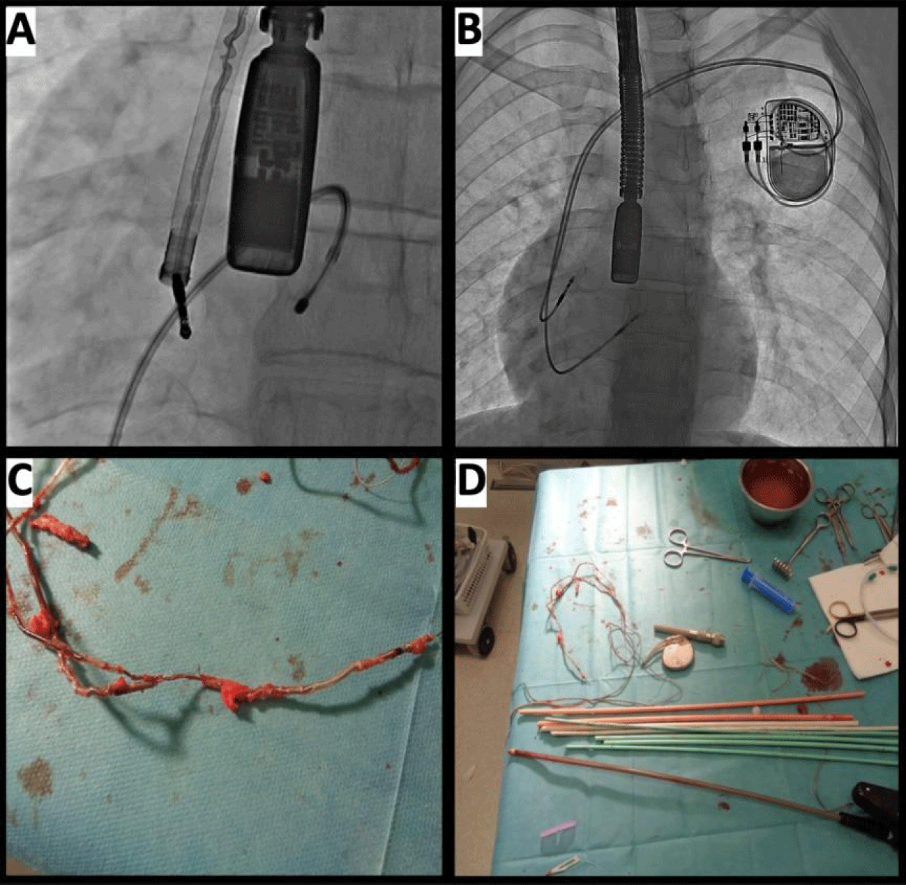
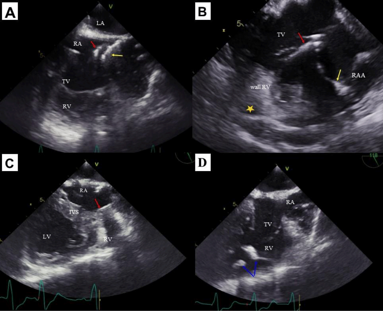
Seventh (rescue) step. In the last stage, there was one more unusual maneuver which brought procedural success. Both electrodes were removed together step by step by one Evolution sheath. The progress was slow, initially the stretching of the right atrium wall was observed until the atrial electrode was removed, and then the classically right ventricular electrode was removed (Figure 2). After the leads had been removed, they still had been connate hard. Finally, it was possible to remove both leads, the two guidewires were via Evolution sheath inserted into the superior vena cava and by the standard introducer the new DDD device was implanted through the regained accesses. The procedure (duration: sheath to sheath time 52 minutes, skin to skin 105 minutes) was successful (Figures 3,4).
The case illustrates technical challenges one may encounter during the subsequent transvenous extraction of implanted in childhood leads. The role of monitoring procedure in aspects visualization of unintentional pooling heart structures cannot be overestimated [30]. Repeated extraction is more difficult but has a similar effectiveness and frequency of adverse events as in the initial procedure [27,28] (Figure 5). Long lead body dwelling time is a known risk factor for developing severe complications during TLE [31,32].
The presence of the complete venous occlusion, incorrect leads position in the cardiac cavities, strong lead-to-lead and lead-to-vein adherence, calcifications and scar tissue that grows over time generates were technical difficulties during TLE, which significantly extended and extremely procedure complexity [21-26]. Improper handling of such technical problems can drive to serious complications [31-35]. The use of atypical techniques, non-standard tricks & tips and TLE dedicated & non-dedicated tools as rescue options in a reference center by an experienced operator proved to be effective and safe. We can consider why didn’t the patient received epicardial pacing leads from the beginning?
Whole medicine, not only electrotherapy, have had in its history different eras. Of course, pacing in children started with epicardial form, too. There were a lot of problems with permanent epicardial pacing in years 1980-90-2000. In this period of time, permanent endocardial pacing go to be more and more popular in children, however there were no steroid eluating endocardial as well as epicardial leads. The literature is full of reports about advantages of endocardial pacing in children [1-6]. We needed many years, to recognize the scale of problems and their meaning when it comes to endocardial pacing in growing child. With child getting older multiple problems arrive throughout juvenescence, such as leads becoming too short, very intensive connecting tissue growth and unfavourable connecting tissue maturation (mineralisation, calcification and even ossification). As a consequence we have to face limited lead life-time and frequent necessity of lead replacement. Described boy received his first pacing system in the era of enthusiasm of endocardial pacing in children.
Conclusion
- Most difficult patient for TLE is adult patient with leads implanted in childhood, young patient with very long pacing history.
- Single lead CIED system seems optimal for children and juveniles in aspect of necessary lead replacement in future.
- The second TLE (re-extraction procedure) may be more complicated than the first one or even by challenging procedure.
- Technical difficulties may prolong the procedure time and make it extremely complicated; we have to avoid major complication but inchoated procedure have to be completed.
- Scope of technical problems is large and in most of cases non-standard tips and tricks & with utility TLE-dedicated tools but in not standard manner may have to be used.
- Rescue options to solve numerous problems should contain the part of TLE education and training.
References
- Rausch CM, Hughes BH, Runciman M, Law IH, Bradley DJ, Sujeev M, Duke A, Schaffer M, Collins KK. Axillary versus infraclavicular placement for endocardial heart rhythm devices in patients with pediatric and congenital heart disease. Am J Cardiol. 2010 Dec 1;106(11):1646-1651. doi: 10.1016/j.amjcard.2010.07.025. Epub 2010 Oct 19. PMID: 21094368.
- Silvetti MS, Drago F, Marcora S, Rav L. Outcome of single-chamber, ventricular pacemakers with transvenous leads implanted in children. Europace. 2007;9:894-899. https://tinyurl.com/4abmzdh4
- Fortescue EB, Berul CI, Cecchin F, Walsh EP, Triedman JK, Alexander ME. Patient, procedural, and hardware factors associated with pacemaker lead failures in pediatrics and congenital heart disease. Heart Rhythm. 2004 Jul;1(2):150-159. doi: 10.1016/j.hrthm.2004.02.020. PMID: 15851146.
- Atallah J, Erickson CC, Cecchin F, Dubin AM, Law IH, Cohen MI, Lapage MJ, Cannon BC, Chun TU, Freedenberg V, Gierdalski M, Berul CI; Pediatric and Congenital Electrophysiology Society (PACES). Multi-institutional study of implantable defibrillator lead performance in children and young adults: results of the Pediatric Lead Extractability and Survival Evaluation (PLEASE) study. Circulation. 2013 Jun 18;127(24):2393-2402. doi: 10.1161/CIRCULATIONAHA.112.001120. Epub 2013 May 21. PMID: 23694966.
- Silvetti MS, Drago F, Grutter G, De Santis A, Di Ciommo V, Ravà L. Twenty years of paediatric cardiac pacing: 515 pacemakers and 480 leads implanted in 292 patients. Europace. 2006 Jul;8(7):530-536. doi: 10.1093/europace/eul062. PMID: 16798767.
- Fortescue EB, Berul CI, Cecchin F, Walsh EP, Triedman JK, Alexander ME. Patient, procedural, and hardware factors associated with pacemaker lead failures in pediatrics and congenital heart disease. Heart Rhythm. 2004 Jul;1(2):150-159. doi: 10.1016/j.hrthm.2004.02.020. PMID: 15851146.
- Lotfy W, Hegazy R, AbdElAziz O, Sobhy R, Hasanein H, Shaltout F. Permanent cardiac pacing in pediatric patients. Pediatr Cardiol. 2013 Feb;34(2):273-280. doi: 10.1007/s00246-012-0433-2. Epub 2012 Aug 12. PMID: 22886361.
- Singh HR, Batra AS, Balaji S. Cardiac pacing and defibrillation in children and young adults. Indian Pacing Electrophysiol J. 2013;13:4-13. https://tinyurl.com/2p9axsuu
- McCanta AC, Schaffer MS, Collins KK. Pediatric and adult congenital endocardial lead extraction or abandonment decision (pacelead) survey of lead management. Pacing Clin Electrophysiol. 2011 Dec;34(12):1621-1627. doi: 10.1111/j.1540-8159.2011.03226.x. Epub 2011 Sep 28. PMID: 21955103.
- Suga C, Hayes DL, Hyberger LK, Lloyd MA. Is there an adverse outcome from abandoned pacing leads? J Interv Card Electrophysiol. 2000 Oct;4(3):493-499. doi: 10.1023/a:1009860514724. PMID: 11046188.
- Silvetti MS, Drago F. Outcome of young patients with abandoned, nonfunctional endocardial leads. Pacing Clin Electrophysiol. 2008;31:473-479. https://tinyurl.com/4txxst2f
- Roux JF, Pagé P, Dubuc M, Thibault B, Guerra PG, Macle L, Roy D, Talajic M, Khairy P. Laser lead extraction: predictors of success and complications. Pacing Clin Electrophysiol. 2007 Feb;30(2):214-220. doi: 10.1111/j.1540-8159.2007.00652.x. PMID: 17338718.
- Lau YR, Gillette PC, Buckles DS, Zeigler VL. Actuarial survival of transvenous pacing leads in a pediatric population. Pacing Clin Electrophysiol. 1993 Jul;16(7 Pt 1):1363-1367. doi: 10.1111/j.1540-8159.1993.tb01729.x. PMID: 7689200.
- Spittell PC, Vlietstra RE, Hayes DL, Higano ST. Venous obstruction due to permanent transvenous pacemaker electrodes: treatment with percutaneous transluminal balloon venoplasty. Pacing Clin Electrophysiol. 1990 Mar;13(3):271-274. doi: 10.1111/j.1540-8159.1990.tb02040.x. PMID: 1690399.
- Figa FH, McCrindle BW, Bigras JL, Hamilton RM, Gow RM. Risk factors for venous obstruction in children with transvenous pacing leads. Pacing Clin Electrophysiol. 1997 Aug;20(8 Pt 1):1902-9. doi: 10.1111/j.1540-8159.1997.tb03594.x. PMID: 9272526.
- Böhm A, Pintér A, Duray G, Lehoczky D, Dudás G, Tomcsányi I, Préda I. Complications due to abandoned noninfected pacemaker leads. Pacing Clin Electrophysiol. 2001 Dec;24(12):1721-1724. doi: 10.1046/j.1460-9592.2001.01721.x. PMID: 11817804.
- Jacheć W, Polewczyk A, Segreti L, Bongiorni MG, Kutarski A. To abandon or not to abandon: Late consequences of pacing and ICD lead abandonment. Pacing Clin Electrophysiol. 2019 Jul;42(7):1006-1017. doi: 10.1111/pace.13715. Epub 2019 May 13. PMID: 31046136.
- Grazia Bongiorni M, Dagres N, Estner H, Pison L, Todd D, Blomstrom-Lundqvist C; Scientific Initiative Committee, European Heart Rhythm Association. Management of malfunctioning and recalled pacemaker and defibrillator leads: Results of the european heart rhythm association survey. Europace. 2014 Nov;16(11):1674-1678. doi: 10.1093/europace/euu302. PMID: 25344962.
- Hussein AA, Tarakji KG, Martin DO, Gadre A, Fraser T, Kim A, Brunner MP, Barakat AF, Saliba WI, Kanj M, Baranowski B, Cantillon D, Niebauer M, Callahan T, Dresing T, Lindsay BD, Gordon S, Wilkoff BL, Wazni OM. Cardiac implantable electronic device infections: added complexity and suboptimal outcomes with previously abandoned leads. JACC Clin Electrophysiol. 2017 Jan;3(1):1-9. doi: 10.1016/j.jacep.2016.06.009. Epub 2016 Sep 7. PMID: 29759687.
- Segreti L, Rinaldi CA, Claridge S, Svendsen JH, Blomstrom-Lundqvist C, Auricchio A, Butter C, Dagres N, Deharo JC, Maggioni AP, Kutarski A, Kennergren C, Laroche C, Kempa M, Magnani A, Casteigt B, Bongiorni MG; ELECTRa Investigators. Procedural outcomes associated with transvenous lead extraction in patients with abandoned leads: an ESC-EHRA ELECTRa (European Lead Extraction ConTRolled) Registry Sub-Analysis. Europace. 2019 Apr 1;21(4):645-654. doi: 10.1093/europace/euy307. PMID: 30624715.
- Cooper JM, Stephenson EA, Berul CI, Walsh EP, Epstein LM. Implantable cardioverter defibrillator lead complications and laser extraction in children and young adults with congenital heart disease: Implications for implantation and management. J Cardiovasc Electrophysiol. 2003;14:344-349. https://tinyurl.com/39yhpmmt
- Friedman RA1, Van Zandt H, Collins E, LeGras M, Perry J. Lead extraction in young patients with and without congenital heart disease using the subclavian approach. Pacing Clin Electrophysiol. 1996;19:778-783. https://tinyurl.com/46c4xzww
- Moak JP, Freedenberg V, Ramwell C, Skeete A. Effectiveness of excimer laser-assisted pacing and ICD lead extraction in children and young adults. Pacing Clin Electrophysiol. 2006 May;29(5):461-466. doi: 10.1111/j.1540-8159.2006.00376.x. PMID: 16689839.
- Zartner PA, Wiebe W, Toussaint-Goetz N, Schneider MB. Lead removal in young patients in view of lifelong pacing. Europace. 2010;12:714-718. https://tinyurl.com/yc57yeuv
- McCanta AC, Tanel RE, Gralla J, Runciman DM, Collins KK. The fate of nontargeted endocardial leads during the extraction of one or more targeted leads in pediatrics and congenital heart disease. Pacing Clin Electrophysiol. 2014 Jan;37(1):104-108. doi: 10.1111/pace.12282. Epub 2013 Oct 25. PMID: 24164671.
- Cecchin F, Atallah J, Walsh EP, Triedman JK, Alexander ME, Berul CI. Lead extraction in pediatric and congenital heart disease patients. Circ Arrhythm Electrophysiol. 2010 Oct;3(5):437-444. doi: 10.1161/CIRCEP.110.957324. Epub 2010 Aug 20. PMID: 20729392.
- Fu H, Ho G, Yang M, Huang X, Fender EA, Mulpuru S, Asirvatham R, Pretorius VG, Friedman PA, Birgersdotter-Green U, Cha YM. Outcomes of repeated transvenous lead extraction. Pacing Clin Electrophysiol. 2018 Oct;41(10):1321-1328. doi: 10.1111/pace.13464. Epub 2018 Aug 23. PMID: 30058073.
- Saeed O, Gupta A, Gross JN, Palma EC. Rate of cardiovascular implantable electronic device (CIED) re-extraction after recurrent infection. Pacing Clin Electrophysiol. 2014 Aug;37(8):963-968. doi: 10.1111/pace.12407. Epub 2014 Apr 26. PMID: 24766634.
- Polewczyk M, Tomków K, Tułecki Ł, Polewczyk A, Mlynarczyk K, Kutarski A. Abandoned pacing lead - a trap and a source of following technical complications during transvenous lead extraction. Heart Beat. 2018;3:90-92. https://tinyurl.com/5e54a4y3
- Nowosielecka D, Polewczyk A, Jacheć W, Kleinrok A, Tułecki Ł, Kutarski A. Transesophageal echocardiography for the monitoring of transvenous lead extraction. Kardiol Pol. 2020 Dec 23;78(12):1206-1214. doi: 10.33963/KP.15651. Epub 2020 Oct 16. PMID: 33078921.
- Jacheć W, Polewczyk A, Polewczyk M, Tomasik A, Kutarski A. Transvenous Lead Extraction SAFeTY Score for Risk Stratification and Proper Patient Selection for Removal Procedures Using Mechanical Tools. J Clin Med. 2020 Jan 28;9(2):361. doi: 10.3390/jcm9020361. PMID: 32013032; PMCID: PMC7073714.
- Jacheć W, Polewczyk A, Polewczyk M, Tomasik A, Janion M, Kutarski A. Risk factors predicting complications of transvenous lead extraction. Biomed Res Int. 2018;8796704. https://tinyurl.com/yn8k9h77
- Kutarski A, Czajkowski M, Pietura R, Obszanski B, Jachec W, Polewczyk M, Mlynarczyk K, Grabowski M, Opolski G. Effectiveness, safety, and long-term outcomes of non-powered mechanical sheaths for transvenous lead extraction. Europace. 2018;20:1324-1333. https://tinyurl.com/bdzj43c4
- Gould J, Sidhu BS, Porter B, Sieniewicz BJ, Teall T, Williams SE, Shetty A, Bosco P, Blauth C, Gill J, Rinaldi CA. Prolonged lead dwell time and lead burden predict bailout transfemoral lead extraction. Pacing Clin Electrophysiol. 2019 Oct;42(10):1355-1364. doi: 10.1111/pace.13791. Epub 2019 Sep 5. PMID: 31433064.
- Zucchelli G, Di Cori A, Segreti L, Laroche C, Blomstrom-Lundqvist C, Kutarski A, Regoli F, Butter C, Defaye P, Pasquié JL, Auricchio A, Maggioni AP, Bongiorni MG; ELECTRa Investigators. Major cardiac and vascular complications after transvenous lead extraction: acute outcome and predictive factors from the ESC-EHRA ELECTRa (European Lead Extraction ConTRolled) registry. Europace. 2019 May 1;21(5):771-780. doi: 10.1093/europace/euy300. PMID: 30590520.
Content Alerts
SignUp to our
Content alerts.
 This work is licensed under a Creative Commons Attribution 4.0 International License.


