Medicine Group . 2022 April 26;3(4):406-407. doi: 10.37871/jbres1456.
Myeloid Sarcoma of the Paranasal Sinus as Solo Manifestation of Acute Myeloid Leukaemia
Andre Carcao*, Ana Isabel Goncalves, Delfim Duarte, Marta Neves and Gustavo Lopes
Introduction
Myeloid Sarcoma (MS) is an extramedullary manifestation of Acute Myeloid Leukemia (AML) but it can be the only manifestation of the disease [1,2]. The disease can involve any body site usually as isolated lesion and involvement of paranasal sinus is rare [2,3]. We report the case of a 73-year-old woman presenting with isolated myeloid sarcoma of the right nasal cavity with no systemic manifestations of disease. Histologic and immunohistochemistry settled the diagnosis of myeloid sarcoma. This kind of cases is frequently misdiagnosed since myeloid sarcoma it is not contemplated as initial differential diagnosis [3,4]. Appropriate recognition and prompt diagnosis is mandatory to ensure early treatment and improve prognosis [3,4].
Case Report
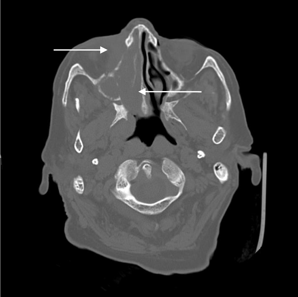
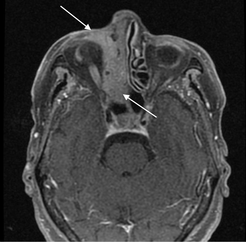
A 73-year-old woman was admitted to the Otorhinolaryngology Emergency Department with complaints of right nasal obstruction associated with nasal swelling. Nasal endoscopic examination detected a whitish homogeneous mass occupying the right nasal cavity. Cervical lymphadenopaty was noted in submandibular region (level Ia) with 3 centimeters, non-tender, firm and painless on palpation. Computed Tomography (CT) and MRI identified a right ethmoidal mass involving the maxillary sinus and the orbit (Figures 1,2). A diagnosis of nasal neoplasm was established and biopsy was performed. Immunohistochemistry examination of the tissue sections revealed diffuse infiltration with high mitotic index, extensively positive for CD34 and CD117. The remaining laboratory tests such as blood count and myelogram did not show any alteration. The positron emission tomography has only showed involvement of the right paranasal region with no other areas being involved. The patient was transferred to the haematology department and the diagnosis of MS was recognised. Treatment with azacitidine and radiation therapy (30 Gy in 15 fractions) were implemented. After 6-month follow-up, the patient is completing the treatment with azacitidine showing no nasal or systemic symptoms.
Discussion
MS of the paranasal sinuses is extremely rare, and its characteristics are not fully understood [5]. MS is mainly associated with AML, and it can be classified into three groups according to the onset of the disease: an extramedullary presentation of acute leukemia, detected simultaneously with the disease; an extramedullary presentation of myelodysplasitc syndromes, chronic myeloid leukemia, or other myeloproliferative diseases; or finally an extramedulary tumor preceding the onset of AML at which the bone marrow aspiration reveals no hematological disease [1]. After the evaluation of our patient we can state that her presentation can be classified as an extramedulary tumour probably preceding the onset of AML.
Interestingly, rates of misdiagnosis of primary MS have been reported in the range of 25-47%, and many primary MS patients are misdiagnosed with lymphoma [5,6]. In previous works, the most useful markers for diagnosis are myeloperoxidase and CD 117 classic myeloid differentiation markers and CD 34 and TdT, markers expressed in immature cells [7,8]. Biopsy of our patient was extensively positive for CD 117 and CD 34, showing typical signs of MS involvement.
While there is no unanimity on the treatment, current options for MS are largely dependent on whether they develop at initial diagnosis or at relapse [2,5]. Concurrent MS with bone marrow involvement is generally treated with chemotherapy directed to the underlying leukemia [1,2,5]. For isolated MS, some reviews have reported that systemic treatment is recommended since there is a higher rate of progression to leukemia [9,10]. High-dose chemotherapy followed by allogeneic transplant of hematopoietic stem cells appears to be a therapeutic option after disease relapse [1,10]. Radiotherapy can be used after initial chemotherapy for better local disease control [10]. In this case, the preferred treatment was systemic chemotherapy with an hypomethylating agent (Azacitidine) followed by radiation therapy given the restrict location of the mass. The allogeneic transplant has not been yet equated for this patient after the good initial response to the treatment.
Conclusion
MS is a rare disease that can be present in a patient without leukaemia and it should be considered in differential diagnosis of nasal neoplasms. Immunohistological investigations combined with clinical features and radiological investigations is mandatory for establishing a correct diagnosis. Early detection and systemic chemotherapy with radiotherapy are vital for better prognosis, as shown in this case.
References
- Yilmaz AF, Saydam G, Sahin F, Baran Y. Granulocytic sarcoma: a systematic review. Am J Blood Res. 2013 Dec 18;3(4):265-70. PMID: 24396704; PMCID: PMC3875275.
- Avni B, Koren-Michowitz M. Myeloid sarcoma: current approach and therapeutic options. Ther Adv Hematol. 2011 Oct;2(5):309-16. doi: 10.1177/2040620711410774. PMID: 23556098; PMCID: PMC3573418.
- Noh BW, Park SW, Chun JE, Kim JH, Kim HJ, Lim MK. Granulocytic Sarcoma in the Head and Neck: CT and MR Imaging Findings. Clin Exp Otorhinolaryngol. 2009 Jun;2(2):66-71. doi: 10.3342/ceo.2009.2.2.66. Epub 2009 Jun 27. PMID: 19565030; PMCID: PMC2702726.
- Neiman RS, Barcos M, Berard C, Bonner H, Mann R, Rydell RE, Bennett JM. Granulocytic sarcoma: a clinicopathologic study of 61 biopsied cases. Cancer. 1981 Sep 15;48(6):1426-37. doi: 10.1002/1097-0142(19810915)48:6<1426::aid-cncr2820480626>3.0.co;2-g. PMID: 7023656.
- Suzuki J, Harazaki Y, Morita S, Kaga Y, Nomura K, Sugawara M, Katori Y. Myeloid Sarcoma of the Paranasal Sinuses in a Patient with Acute Myeloid Leukemia. Tohoku J Exp Med. 2018 Oct;246(2):141-146. doi: 10.1620/tjem.246.141. PMID: 30369515.
- Meis JM, Butler JJ, Osborne BM, Manning JT. Granulocytic sarcoma in nonleukemic patients. Cancer. 1986 Dec 15;58(12):2697-709. doi: 10.1002/1097-0142(19861215)58:12<2697::aid-cncr2820581225>3.0.co;2-r. PMID: 3465429.
- Yamauchi K, Yasuda M. Comparison in treatments of nonleukemic granulocytic sarcoma: report of two cases and a review of 72 cases in the literature. Cancer. 2002 Mar 15;94(6):1739-46. doi: 10.1002/cncr.10399. PMID: 11920536.
- Raphael J, Valent A, Hanna C, Auger N, Casiraghi O, Ribrag V, De Botton S, Saada V. Myeloid sarcoma of the nasopharynx mimicking an aggressive lymphoma. Head Neck Pathol. 2014 Jun;8(2):234-8. doi: 10.1007/s12105-013-0495-3.
- Yilmaz AF, Saydam G, Sahin F, Baran Y. Granulocytic sarcoma: a systematic review. Am J Blood Res. 2013 Dec 18;3(4):265-70. PMID: 24396704; PMCID: PMC3875275.
- Avni B, Koren-Michowitz M. Myeloid sarcoma: current approach and therapeutic options. Ther Adv Hematol. 2011 Oct;2(5):309-16. doi: 10.1177/2040620711410774. PMID: 23556098; PMCID: PMC3573418.
- Noh BW, Park SW, Chun JE, Kim JH, Kim HJ, Lim MK. Granulocytic Sarcoma in the Head and Neck: CT and MR Imaging Findings. Clin Exp Otorhinolaryngol. 2009 Jun;2(2):66-71. doi: 10.3342/ceo.2009.2.2.66. Epub 2009 Jun 27. PMID: 19565030; PMCID: PMC2702726.
- Neiman RS, Barcos M, Berard C, Bonner H, Mann R, Rydell RE, Bennett JM. Granulocytic sarcoma: a clinicopathologic study of 61 biopsied cases. Cancer. 1981 Sep 15;48(6):1426-37. doi: 10.1002/1097-0142(19810915)48:6<1426::aid-cncr2820480626>3.0.co;2-g. PMID: 7023656.
- Suzuki J, Harazaki Y, Morita S, Kaga Y, Nomura K, Sugawara M, Katori Y. Myeloid Sarcoma of the Paranasal Sinuses in a Patient with Acute Myeloid Leukemia. Tohoku J Exp Med. 2018 Oct;246(2):141-146. doi: 10.1620/tjem.246.141. PMID: 30369515.
- Meis JM, Butler JJ, Osborne BM, Manning JT. Granulocytic sarcoma in nonleukemic patients. Cancer. 1986 Dec 15;58(12):2697-709. doi: 10.1002/1097-0142(19861215)58:12<2697::aid-cncr2820581225>3.0.co;2-r. PMID: 3465429.
- Yamauchi K, Yasuda M. Comparison in treatments of nonleukemic granulocytic sarcoma: report of two cases and a review of 72 cases in the literature. Cancer. 2002 Mar 15;94(6):1739-46. doi: 10.1002/cncr.10399. PMID: 11920536.
- Raphael J, Valent A, Hanna C, Auger N, Casiraghi O, Ribrag V, De Botton S, Saada V. Myeloid sarcoma of the nasopharynx mimicking an aggressive lymphoma. Head Neck Pathol. 2014 Jun;8(2):234-8. doi: 10.1007/s12105-013-0495-3. Epub 2013 Oct 8. PMID: 24101200; PMCID: PMC4022928.
- Pileri SA, Ascani S, Cox MC, Campidelli C, Bacci F, Piccioli M, Piccaluga PP, Agostinelli C, Asioli S, Novero D, Bisceglia M, Ponzoni M, Gentile A, Rinaldi P, Franco V, Vincelli D, Pileri A Jr, Gasbarra R, Falini B, Zinzani PL, Baccarani M. Myeloid sarcoma: clinico-pathologic, phenotypic and cytogenetic analysis of 92 adult patients. Leukemia. 2007 Feb;21(2):340-50. doi: 10.1038/sj.leu.2404491. Epub 2006 Dec 14. PMID: 17170724.
- Avni B, Rund D, Levin M, Grisariu S, Ben-Yehuda D, Bar-Cohen S, Paltiel O. Clinical implications of acute myeloid leukemia presenting as myeloid sarcoma. Hematol Oncol. 2012 Mar;30(1):34-40. doi: 10.1002/hon.994. Epub 2011 Jun 2. PMID: 21638303.
- Epub 2013 Oct 8. PMID: 24101200; PMCID: PMC4022928.
- Pileri SA, Ascani S, Cox MC, Campidelli C, Bacci F, Piccioli M, Piccaluga PP, Agostinelli C, Asioli S, Novero D, Bisceglia M, Ponzoni M, Gentile A, Rinaldi P, Franco V, Vincelli D, Pileri A Jr, Gasbarra R, Falini B, Zinzani PL, Baccarani M. Myeloid sarcoma: clinico-pathologic, phenotypic and cytogenetic analysis of 92 adult patients. Leukemia. 2007 Feb;21(2):340-50. doi: 10.1038/sj.leu.2404491. Epub 2006 Dec 14. PMID: 17170724.
- Avni B, Rund D, Levin M, Grisariu S, Ben-Yehuda D, Bar-Cohen S, Paltiel O. Clinical implications of acute myeloid leukemia presenting as myeloid sarcoma. Hematol Oncol. 2012 Mar;30(1):34-40. doi: 10.1002/hon.994. Epub 2011 Jun 2. PMID: 21638303.
Content Alerts
SignUp to our
Content alerts.
 This work is licensed under a Creative Commons Attribution 4.0 International License.


