Medicine Group . 2022 June 24;3(6):712-713. doi: 10.37871/jbres1501.
Type II and IV Thumb Duplication in Neonates
Adil Khan1, Briana Hernandez2, Justin Harrell3 and Shabih Manzar4*
2Intern, Department of Pediatrics, Louisiana State University Health Sciences Center, Shreveport, LA, USA
3Resident, Department of Medicine-Pediatrics, Louisiana State University Health Sciences Center, Shreveport, LA, USA
4Attending, Department of Pediatrics, Louisiana State University Health Sciences Center, Shreveport, LA, USAv
Summary
Thumb duplication is a rare anomaly. Here we describe two neonatal cases of thumb duplications, one type II and the other type IV. Seven types have been described in the medical literature. Radiological assessment is important in differentiating between these types.
Case 1
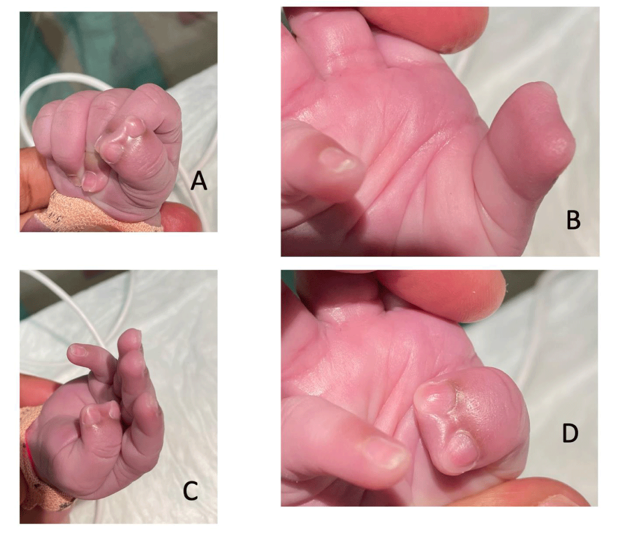
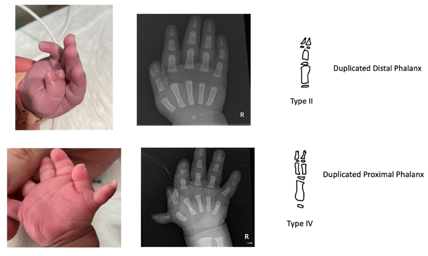
A male infant was born at term. On careful physical examination, he was noted to have an isolated anomaly of the right thumb (Figure 1A-D).
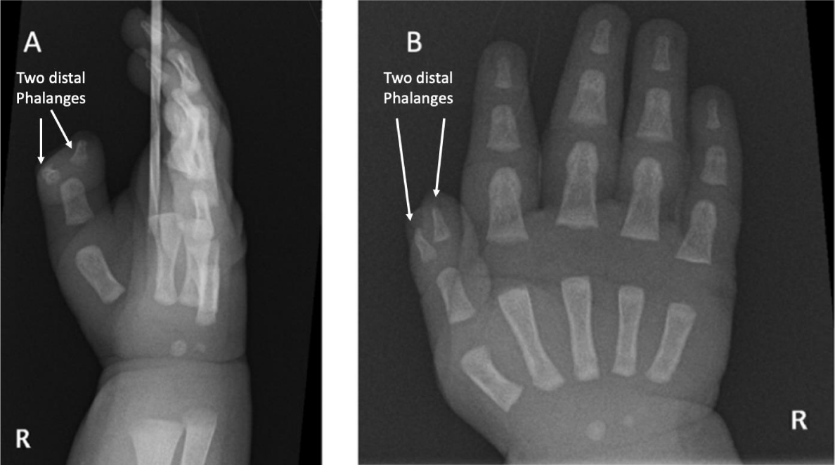
The infant had no dysmorphic features, and the remainder of the physical examination was within normal limits. Mother reported no family history of similar problems among close or distant families on both maternal and paternal sides. Radiological evaluation was performed which showed complete duplication of the distal phalanx of the thumb (Figure 2A,B).
Case 2
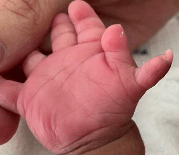
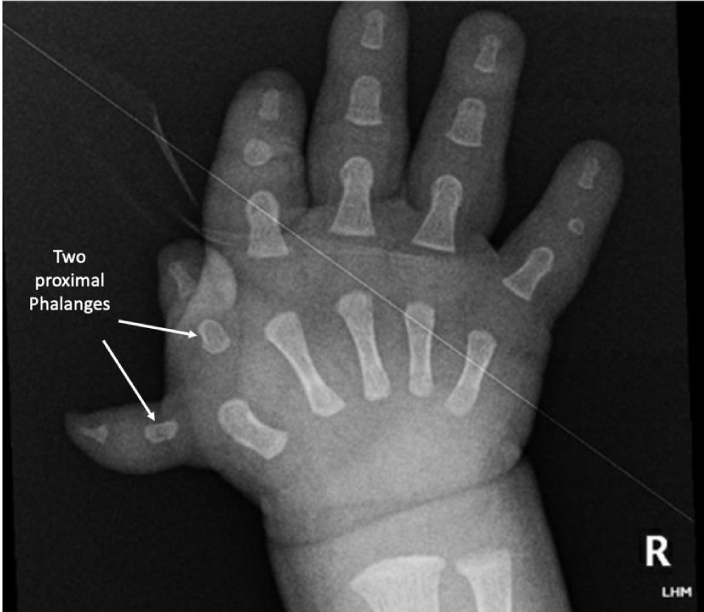
An infant was born with clinical features of Down syndrome. The chromosomal analysis confirmed the diagnosis of Trisomy 21 (47, XY, +21). A picture of the newborn’s hand is shown below (Figure 3A,B).
| Table 1: Classification of thumb duplication (Watt AJ [2]) | ||
| Wassel Type | Anatomic Description | Percentage |
| Type 1 | Bifid Distal Phalanx | 4% |
| Type II | Duplicated Distal Phalanx | 16% |
| Type III | Bifid Proximal Phalanx | 11% |
| Type IV | Duplicated Proximal Phalanx | 40% |
| Type V | Bifid Metacarpal | 10% |
| Type VI | Duplicated Metacarpal | 4% |
| Type VII | Triphalangeal Thumb | |
Discussion
The infant described in case 1 had type II pre-axial thumb duplication (two distal phalanges), while case 2 had a polydactyly, classified as type IV pre-axial thumb duplication (two proximal phalanges). Post-axial polydactyly would be on the ulnar side of the hand.
Thumb duplication is a rare anomaly; the incidence is 1 in every 3,000 live births, with the highest incidence among Asian and Caucasian populations [1]. Seven types (I-VII) of thumb anomalies have been described in literature [2].
These cases highlighted the need for obtaining radiological studies to differentiate between different types of thumb duplication. Both infants were referred to hand surgery team for further intervention. Surgical treatment for congenital thumb duplication ranges from simple ablation to a reconstruction. The aim is to correct any angular deformity by rebalancing of the soft tissue with collateral ligament reconstruction and tendon centralization. In cases of osseous deformity that disrupt the axial alignment of the thumb, closing or opening wedge osteotomies may be necessary to provide alignment followed by bone graft, obtained from the amputated thumb [2].
References
- Townsend DJ, Lipp EB Jr, Chun K, Reinker K, Tuch B. Thumb duplication, 66 years' experience--a review of surgical complications. J Hand Surg Am. 1994 Nov;19(6):973-6. doi: 10.1016/0363-5023(94)90099-X. PMID: 7876498.
- Watt AJ, Chung KC. Duplication. Hand Clin. 2009 May;25(2):215-27. doi: 10.1016/j.hcl.2009.01.001. PMID: 19380061.
Content Alerts
SignUp to our
Content alerts.
 This work is licensed under a Creative Commons Attribution 4.0 International License.


