Medicine Group . 2022 June 30;3(6):729-733. doi: 10.37871/jbres1505.
A Novel Approach to the Respiratory Disease Follow-up Based on Plethhysmography Signal Parameters
Rene I Gonzalez Fernandez*, Jose L Hernandez Caceres and Jorge G Perez Blanco
- Respiratory disease follow-up
- Pulse oximetry
- PPG derived respiratory rate
- Chronic obstructive pulmonary disease
Abstract
Aim: The aim of this paper is to present a novel approach for the monitoring of respiratory diseases based on the combined study of heart rate, respiratory rate and Pulse Oximetry (SpO2).
Introduction: Since the start of the COVID-19 pandemic, respiratory diseases have increased its worldwide prevalence dramatically. Added to the impact of the pandemic is the already existing situation of a growing prevalence of diseases such as bronchial asthma and Chronic Obstructive Pulmonary Disease (COPD). On the other hand, it is known that the cardiovascular system is overloaded when there are respiratory insufficiencies and this condition can cause severe damage to health. Pulmonology and Cardiology are independent medical specialties that deal with these diseases and often do not cooperate with each other strongly, so a proposal is presented that combines respiratory and cardiovascular variables to deal with this health problem.
Methods: A prototype was developed based on the STM32L073CZTx processor controlling the E305654 pulse oximetry module, which on demand delivers SpO2, photo plethysmography signal samples and pulse rate values. A vector was computed, every thirty minutes, to combine the respiratory rate obtained from the Plethysmography Signal (PPG), the mean value of SpO2 and the pulse rate. The vector module represents the cardiovascular and respiratory systems performance.
Results: Twenty healthy volunteers, twenty people with COPD and five subjects suffering bronchial asthma, during periods without crisis, were studied. The dispersion within the group of each patient was not significant, but a notable difference can be observed between the healthy volunteers and the other people studied. A "normal region" can be set with clearly defined borders.
Conclusion: The proposed solution seems promising for evaluating respiratory function even when it is compensated by a cardiac response, making it easier to identify people suffering from respiratory diseases and who may appear normal or healthy.
Abbreviations
PPG: Photoplethysmography; RR: Respiratory Rate; HR: Heart Rate
Introduction
Acute respiratory diseases have a growing impact on the worldwide morbidity and mortality. According to data from the World Health Organization (WHO), there are 235 million people suffering asthma and 64 million with Chronic Obstructive Pulmonary Disease (COPD) [1]. Today, the impact of COVID-19 in all countries has dramatically increased the number of people treated for respiratory disorders [2,3]. It is usual that while suffering from the disease or in the post-disease period, severe complications could be lethal [3].
It is well known that the respiratory and cardiovascular systems work in strongly coordination to transport oxygen throughout the body as a primary element for metabolism. Blood oxygenation tends to decrease in persons suffering respiratory disorders; this deficiency is compensated automatically by an increase in respiratory rate and heart rate. That is why scientists assume that human organism tries to cover respiratory deficiencies with a cardiac overload in order to supply the required oxygen. This cardiorespiratory compensation masks the respiratory deficit and allows this condition to evolve towards more serious states because the respiratory deficiency is not solved and the heart is overloaded. Based on the previous explanation, it is clear why cardiorespiratory function must be studied comprehensively, with Photo Plethysmography (PPG) as the best non-invasive option to achieve this goal. The required instrumentation and connections to the patient are minimized while sufficient information is collected for the intended analysis [4,5].
Respiratory Rate (RR) is studied at different levels of health systems with different objectives. This parameter can be a highly sensitive indicator of an acute deterioration in the health status of the patient. Apart from indicating respiratory deficiency, it is a predictor of severe cardiac disorders due to the close relationship between the respiratory and cardiovascular systems. On the other hand, at the primary health level, the RR study is used for the diagnosis of pneumonia and sepsis, hence its importance.
Pulse Rate (PR) indicates how many times the heart beats per minute, hence its importance in the cardiovascular system study. It is also important in the respiratory system analysis because many times the human organism tends to compensate for respiratory insufficiencies increasing pulse rate and this generates a cardiovascular overload that can lead to subsequent cardiac disorders.
Pulse oximetry (SpO2) allows estimating the arterial blood oxygenation percentage and is therefore a vital parameter for any human body study. When there are respiratory disorders, less oxygen is transported by arterial blood and metabolism is affected, so human automatism mechanisms increase heart rate to pump more blood and transport the requires oxygen.
The combined study of SpO2, pulse rate and heart rate values could be a powerful tool for a comprehensive analysis of the functioning of the cardiovascular. A "healthy space" can be set in a tridimensional axis system to set the borders among normal and abnormal functioning of the cardiorespiratory system. The implementation of this approach can be a low-cost solution because it can be based on a pulse oximetry module controlled by a general-purpose microcontroller, two low-price items nowadays. A quantitative evaluation can be done and a follow-up is easy to set.
The aim of this paper is to discuss the approach described previously. A reliable, comfortable and low-cost follow-up process can be implemented for patients suffering from acute respiratory diseases such as COPD, bronchial asthma or complications of the COVID-19 disease, caused by the SARS-CoV-2 virus.
Materials and Methods
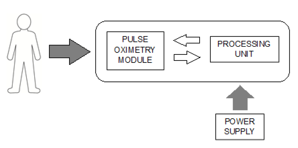
The proposed system is composed of the Recorder and the Analyzer. The Recorder is a medical device that allows the acquisition of II from the standard ECG and two channels of pulse oximetry information simultaneously. A Bluetooth channel is enabled for data transmission between both parts of the system as shown in figure 1. A pairing process between the Recorder and the mobile device running the Analyzer is a mandatory for this type of wireless communication.
The proposed approach for monitoring acute respiratory diseases is based on the acquisition of the PPG signal and the extraction from it of the following parameters: Pulse Rate (PR), Respiratory Rate (RR) and periodic values of oxygen saturation (SpO2). With these variables, a three-dimensional space is built in order to study the patient evolution; it is expected that a region of normality can be set.
The variables mentioned above can be get implementing a low-cost electronic solution and the properly software. The authors developed a prototype composed by the following items:
- An embedded pulse oximetry module, model E305654, from the Chinese company Mind ray.
- A processing unit based on the STM32L073CZTx microcontroller embedding memory, interrupt management, I/O lines, communication and other capabilities.
- A baseboard to implement voltage supply and connection between pulse oximetry module and processing board.
- A medical grade power supply to guarantee the safety of the patients and conversion for alter current to direct current to get the needed voltages.
The block diagram shown in figure 1 represents the design of the developed prototype. On the other hand, embedded software was developed to control the prototype operation, to implement the necessary methods to compute the studied variables and to set communication with a personal computer. This software was programmed in C++ language.
The E305654 oximetry module has a serial interface that allowing getting PPG signal samples and values of SpO2 and PR on request. The PPG sampling rate was set to 250 Hz; SpO2 and PR values were read every ten seconds, obtaining an average value minute by minute.
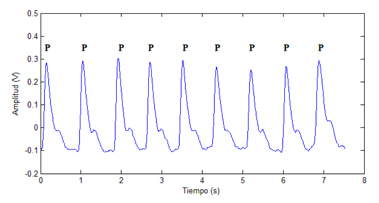
Also, PR was computed using an algorithm developed by the authors and taken as Golden Rule the values read from the oximetry module. It was done for a future implementation in a smart wrist device in development. Peak identification on PPG signal is decisive for PR computing, since the distance between them is the basis for this calculation. Figure 2 shows the PPG signal and the peaks marked with the letter P.
A derivative function, as shown in expression 1, is calculated for peak identification. The zero crossings of this function allow the peaks identification since on both sides of these there must be derivative function values of opposite polarity.
where:
deriv(i): derivative function value.
x(i): PPG signal sample.
Every ten seconds, the average PR interval is calculated and rate is computed using expression 2.
where:
PR: Pulse Rate.
API: Average Pulse Interval for ten seconds.
Multiple algorithms have been proposed in the specialized literature to obtain the RR from the ECG or PPG [6-8]. Generally, these algorithms are based on the Fast Fourier Transform (FFT) and other spectral procedures that represent a considerable processing load for microcontrollers [xx]. Other authors have used wavelet decomposition and digital filtering strategies that minimize the error, reporting an error of less than 0.5 breaths per minute [9], although better results have been obtained with autoregressive approaches [10].
Madhav [10] proposed a method based on the Empirical Mode Decomposition (EMD) for RR estimation based on PPG signal peak to peak series. EDM allows extracting signals from a noisy non-linear and non-stationary process. This ability is based on the identification of oscillatory modes at different time scales. The EMD is an iterative process based on local maximum and minimum peaks identification, creating of corresponding upper and lower envelopes of the studied signal, computing of a mean signal form the envelopes, subtraction this mean signal from the studied signal and a repetition of these steps until the different between the studied signal and the computed mean signal is not significant. The authors preferred to stop at the first loop, the mean envelope calculation, as proposed Fusco [11]. This improvement reduces the algorithm complexity and the computational overload. RR estimation is based on the mean envelope frequency analysis. The Power Spectral Density (PSD) of the respiratory component is used to evaluate the dominant frequency peak that is associated with RR. The exposed approach was tested with from PhysioBank MIMIC II Waveform Database [12], and a good performance was revealed in estimating RR values from PPG signal. The reported Mean Absolute Error (MAE) was 0.0027 Hz, corresponding to 0.19 breaths per minute, and the Spearman’s correlation coefficient was 0.962.
The RR and PR values are scaled percentagewise to their maximum values so that their representation is homogeneous. 40 breaths/minute was considered as the maximum value for RR and 220 beats/minute for PR, since values higher than these are uncommon at rest state. SpO2 is a percentage value and does not require transformation. In this way, the analysis of three-dimensional graphics of these parameters is more evident.
As previously stated, RR, PR and SpO2 values are obtained every 10 minutes and averaged for each hour. Important differences between healthy people and others, suffering respiratory diseases, can be observed representing the results graphically or on tables. The results are transmitted to a personal computer to facilitate their representation and analysis.
Results and Discussion
A prototype of the proposed solution has been developed and tested at laboratory level successfully. Tests were done following the IEC 60601-1 general safety standard for medical devices. The OEM pulse oximetry module and the medical grade power supply are supported by international certifications, such as CE mark, guarantying compliance with current highest quality and safety standards, so patient safety is guaranteed in the proposed solution.
Taking in count the international medical community criteria, a healthy region was set using the following values as frontiers: SpO2 from 96% to 100%; RR from 12 to 18 breaths per minute and HR from 60 to 100 beats per minute. The first remarkable outcome is that all the values recorded from healthy volunteers keep inside the region defined as healthy. For these people, HR was always between 70 and 90 beats per minute, the SpO2 values were between 97% and 99% and respiratory rate between 13 and 16 breaths per minute. However, it should be noted that the values corresponding to the studied asthmatic patients, without crisis and under long-term medication, were also within this healthy region in approximately 40% of the measurements made and the rest were above the normality limits, but very close. A clear difference between asthmatics and healthy people cannot be set, but it is expected that parameter values from asthmatics tend to move far from healthy region when a bigger sample of asthmatic sample be available. COPD patients have a significant different performance; they have always two parameters out of healthy region at least.
Table 1 clearly shows the difference between healthy people and people with COPD. While healthy people achieve SpO2 values above 97% with a normal cardiorespiratory function, in people suffering COPD, heart rate and respiratory rate increase markedly to achieve much lower SpO2 values. This difference can be translated as a dangerous cardiorespiratory overload; it could be the origin of several cardiac disturbances such as ventricular hypertrophy and arrhythmias. A graphic implementation of these values and differences can be a useful tool for an inexperienced medical staff, a very useful feature for primary health level screening purposes.
| Table 1: Twenty measures from healthy and COPD subjects. | ||||||
| Healthy | COPD | |||||
| RR | HR | SpO2 | RR | HR | SpO2 | |
| 1 | 14 | 80 | 96 | 18 | 97 | 94 |
| 2 | 13 | 84 | 97 | 18 | 98 | 94 |
| 3 | 14 | 84 | 98 | 20 | 101 | 93 |
| 4 | 13 | 84 | 98 | 20 | 97 | 94 |
| 5 | 13 | 85 | 96 | 18 | 90 | 93 |
| 6 | 13 | 80 | 98 | 20 | 100 | 93 |
| 7 | 13 | 81 | 97 | 19 | 105 | 93 |
| 8 | 14 | 82 | 97 | 18 | 103 | 93 |
| 9 | 15 | 84 | 98 | 20 | 103 | 93 |
| 10 | 14 | 85 | 96 | 22 | 100 | 93 |
| 11 | 14 | 78 | 98 | 22 | 98 | 94 |
| 12 | 14 | 84 | 98 | 20 | 99 | 94 |
| 13 | 14 | 85 | 98 | 20 | 101 | 92 |
| 14 | 14 | 78 | 98 | 18 | 98 | 94 |
| 15 | 14 | 84 | 97 | 19 | 98 | 93 |
| 16 | 14 | 84 | 98 | 21 | 98 | 94 |
| 17 | 14 | 85 | 98 | 19 | 101 | 93 |
| 18 | 16 | 84 | 97 | 19 | 103 | 94 |
| 19 | 15 | 84 | 98 | 22 | 102 | 93 |
| 20 | 15 | 84 | 97 | 18 | 100 | 93 |
| Mean | 14.00 | 82.95 | 97.40 | 19.55 | 99.60 | 93.35 |
The same difference cannot be observed between the studied healthy people and asthmatics, although the latter tend to parameter´s values very close to the upper limits of the healthy region. Asthmatic subjects under long-term could be near to healthy region when they are without crisis, but their parameters could be near the normal borderline depending of the effectiveness of their medication. This fact could convert the proposed solution in a useful tool to follow-up the emergency attention of asthmatic patients.
Conclusion
The proposed solution seems to be a useful tool to study people suffering from respiratory diseases. Low cost and simplicity are significant features of the proposed solution, there are low-priced OEM pulse oximetry modules on the market and the same goes for microcontroller-based processing units and medical grade power supply blocks. The combined study of respiratory and cardiac variables does not allow respiratory deficit can be masked by a cardiovascular overload without this condition is revealed. Due to its simplicity and low cost, the proposed solution can be extended to the respiratory diseases screening, being ideal for its implementation in low-income countries. More tests are needed, but the results obtained to date are very promising. Heart Rate Variability (HRV) could be a valuable contribution to this approach because it is able to reflect the status of the Autonomous Nervous System (ANS).
Acknowledgement
The authors wish to thank the economic support of the Ministry of Science of the Republic of Cuba for the successful development of this project. In addition, they want to highlight the support provided by the colleagues of the Cuban Center for Neurosciences who contributed their experience and technical criteria to obtain the solutions described in this paper.
References
- World Health Organization. The world health report. 2020.
- Zhu N, Zhang D, Wang W, Li X, Yang B, Song J, Zhao X, Huang B, Shi W, Lu R, Niu P, Zhan F, Ma X, Wang D, Xu W, Wu G, Gao GF, Tan W, China Novel Coronavirus Investigating and Research Team. A novel coronavirus from patients with pneumonia in china, 2019. N Engl J Med. 2020 Feb 20;382(8):727-733. doi: 10.1056/NEJMoa2001017. Epub 2020 Jan 24. PMID: 31978945; PMCID: PMC7092803.
- Carlos WG, Dela Cruz CS, Cao B, Pasnick S, Jamil S. Novel wuhan (2019-nCoV) coronavirus. Am J Respir Crit Care Med. 2020 Feb 15;201(4):P7-P8. doi: 10.1164/rccm.2014P7. PMID: 32004066.
- Sepp VP, Vinson J, Lahtinen O, Hyttinen J. Assessment of breathing parameters during running with a wearable bio impedance device. 4th European Conference of the International Federation for Medical and Biological Engineering. Springer Berlin Heidelberg. 2009.
- Karlen W, Raman S, Ansermino JM, Dumont GA. Multiparameter respiratory rate estimation from the photo plethysmogram. IEEE Trans Biomed Eng. 2013 Jul;60(7):1946-1953. doi: 10.1109/TBME.2013.2246160. Epub 2013 Feb 8. PMID: 23399950.
- Leonard PA, Douglas JG, Grubb NR, Clifton D, Addison PS, Watson JN. A fully automated algorithm for the determination of respiratory rate from the photo plethysmogram. J Clin Monit Comput. 2006 Feb;20(1):33-36. doi: 10.1007/s10877-005-9007-7. Epub 2006 Mar 11. PMID: 16532280.
- Charlton PH, Villarroel M, Salguiero F. Waveform analysis to estimate respiratory Rate. 2016 Sep 10. In: MIT Critical Data, editor. Secondary analysis of electronic health records [Internet]. Cham (CH): Springer; 2016. Chapter 26. PMID: 31314269.
- Charlton PH, Bonnici T, Tarassenko L, Alastruey J, Clifton DA, Beale R, Watkinson PJ. Extraction of respiratory signals from the electrocardiogram and photo plethysmogram: technical and physiological determinants. Physiol Meas. 2017 May;38(5):669-690. doi: 10.1088/1361-6579/aa670e. Epub 2017 Mar 15. PMID: 28296645.
- Meredith DJ, Clifton D, Charlton P, Brooks J, Pugh CW, Tarassenko L. Photo plethysmographic derivation of respiratory rate: A review of relevant physiology. J Med Eng Technol. 2012 Jan;36(1):1-7. doi: 10.3109/03091902.2011.638965. Epub 2011 Dec 20. PMID: 22185462.
- Madhav KV, Ram MR, Krishna EH, Komalla NR, Reddy KA. Estimation of respiration rate from ECG, BP and PPG signals using empirical mode decomposition. Instrumentation and Measurement Technology Conference. 2011.
- Fusco D, Locatelli F, Onorati GC, Durelli, Santambrogio. On how to extract breathing rate from PPG signal using wearable devices. 2015 IEEE Biomedical Circuits and Systems Conference (BioCAS). 2015:1-4. doi: 10.1109/BioCAS.2015.7348369.
- Goldberger AL, Amaral LA, Glass L, Hausdorff JM, Ivanov PC, Mark RG, Mietus JE, Moody GB, Peng CK, Stanley HE. Physiobank, physiotoolkit, and physionet: Components of a new research resource for complex physiologic signals. Circulation. 2000 Jun 13;101(23):E215-220. doi: 10.1161/01.cir.101.23.e215. PMID: 10851218.
Content Alerts
SignUp to our
Content alerts.
 This work is licensed under a Creative Commons Attribution 4.0 International License.


