Medicine Group . 2022 October 31;3(10):1290-1292. doi: 10.37871/jbres1594.
Alveolar Adenoma of the Lung: A Case Report
Rayyan Daghistani1, Reem Alwasiah1*, Hassan AL-Omaish1, Talal Yahyan1 and Amna Almutrafi2
2Department of Pathology and Laboratory Medicine, King Abdulaziz Medical City, National Guard Affairs Hospital, Riyadh, Saudi Arabia
- Biopsy
- Lung
- Hemorrhage
Abstract
Background: An alveolar adenoma is a benign lung lesion that presents as a solitary pulmonary nodule. Only a few reports have been published on this disease, which is typically diagnosed by wedge resection.
Case presentation: We present a 39 year old woman with multiple co-morbidities with an incidental pulmonary alveolar adenoma that was diagnosed utilizing a CT-guided biopsy, she then suffered a rare complication necessitating ICU admission.
Conclusion: We report the rare case of a diagnosis based on a Computed Tomography (CT)-guided biopsy.
Background
Alveolar Adenomas (AAs) are rare benign lung lesions that present themselves as cystic or solid masses. AAs were first reported in 1986 [1], and only 40 reports have been published on this disease since then. According to the Fleischner Society, follow-ups are indicated in patients with these nodules because they are mostly incidental. The only curative treatment is a surgical wedge resection procedure with a postoperative histopathological diagnosis [2,3]. Wang, et al. [4] reported a case of a multifocal cystic mass that was diagnosed as AA based on biopsy findings. This study reports a case of an AA that was initially presented as a cyst and was diagnosed by Computed Tomography (CT)-guided biopsy.
Case Presentation
The patient was a 39-year-old woman with a history of multiple chronic gastrointestinal abnormalities with bowel fistulas, prior surgical history of multiple fistulectomies and anastomoses complicated by short bowel syndrome. The patient was investigated for Crohn’s disease at the time, and results were negative. She was also diagnosed with hypothyroidism and was receiving levothyroxine. The patient experienced hearing loss and had a history of deep-vein thrombosis.
The patient was admitted through the Emergency Room (ER) after she presented diarrhea symptoms (7–10 times/day), which lasted for 1 month, chest pain, and generalized body aches. Upon examination, the patient was alert, oriented, and vitally stable. Her chest was clear, and her abdominal examination was unremarkable.
Laboratory tests revealed electrolyte imbalances: hypokalemia (2.4 mmol/L; normal values 3.5-5.1 mmol/L), hypomagnesemia (0.36 mmol/L; normal values 0.66-1.07 mmol/L), and hypocalcemia (1.77 mmol/L; normal values 2.10-2.55 mmol/L). Accordingly, she was admitted for medical observation and correction.
Discussion
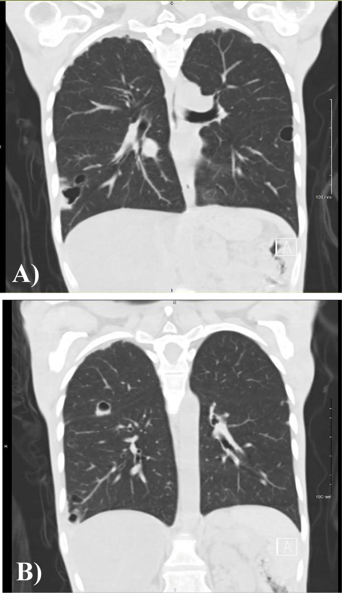
The first CT scan was performed in October 2021 and revealed multiple lung cysts without a particular pattern. The diagnosis was indeterminate cysts, and the proposed differential diagnosis was based on postinfectious pneumatoceles (Figures 1A& 1B).
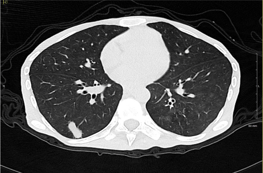
The CT performed in February 2022 (Figure 2) showed opacification of the largest cyst in the right lower lung lobe suggestive of an autoimmune disease. However, the patient’s results were negative for Anti-Neutrophil Cytoplasmic Antibodies (ANCA) and rheumatoid factor. The primary team decided to perform a biopsy of the lesion to establish a definitive diagnosis.
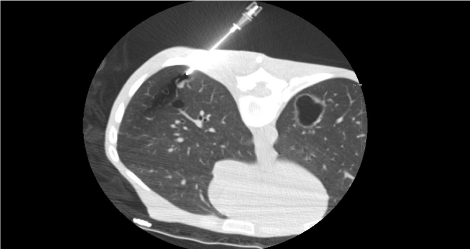
The primary medical team decided to perform a biopsy of the lesion to establish a definitive diagnosis. Another CT was performed later that month when the patient underwent a CT-guided biopsy procedure, which targeted the solid nodule with the use of a 17G coaxial system. Four good cores were successfully acquired (Figure 3).
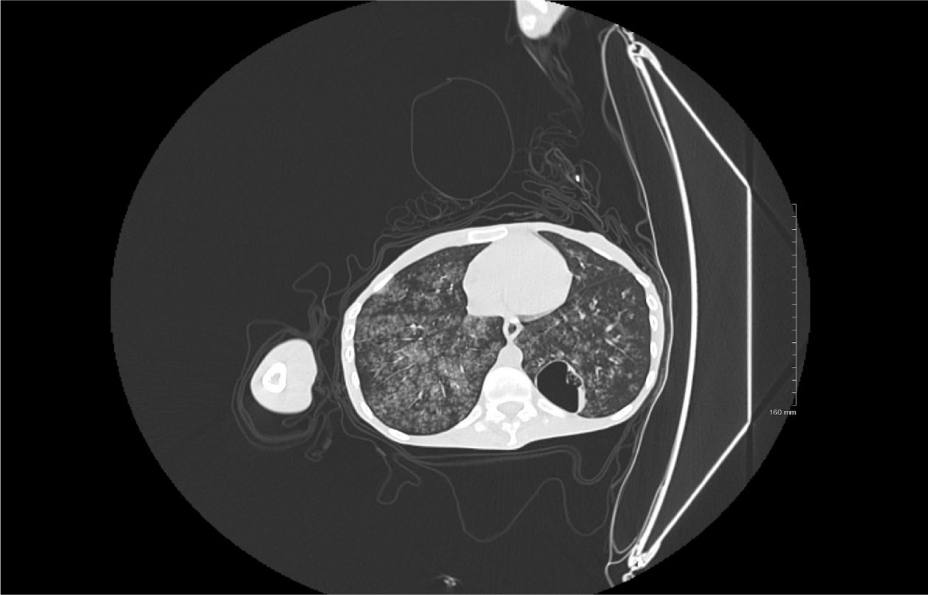
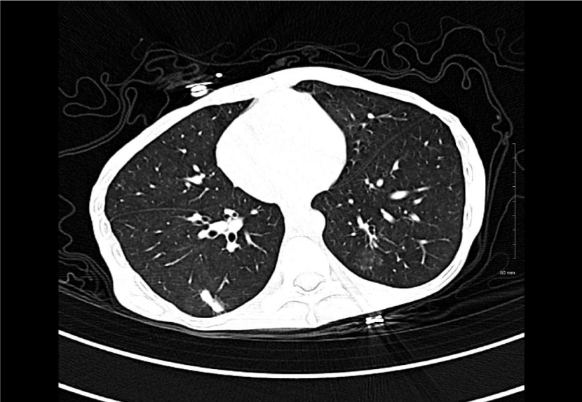
Shortly after the procedure, the patient developed hemoptysis and became unstable. Another CT scan was conducted soon thereafter which did not reveal pneumothorax. However, bilateral ground glass opacities were observed. Owing to the presence of hemoptysis, the findings were associated with alveolar hemorrhage that was likely secondary to aspiration. The patient had a normal coagulation profile (Figure 4).
Local pulmonary hemorrhage is the second most common postbiopsy complication after pneumothorax [5]. There have been no previous reports on bilateral alveolar hemorrhage following biopsy procedures. However, this patient was immediately transferred to the intensive care unit and was managed accordingly.
The follow-up CT scan (performed in March) revealed ground glass opacities with a decrease in the size of the pulmonary nodule (Figure 5).
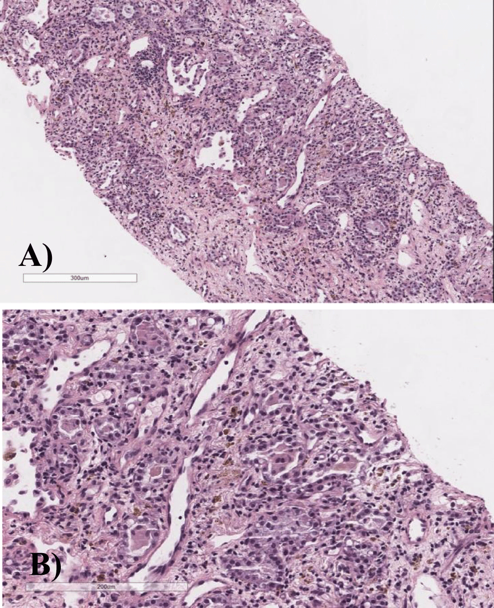
Histological examination revealed a well-circumscribed lesion composed of air spaces, filled with eosinophilic material, and lined with cuboidal bland cells. In immunohistochemistry, the cells were positive for thyroid transcription factor 1. Focal collagen fibers, spindle-shaped cells, and hemosiderin pigments were observed in the interstitium. Overall, the histological findings supported the diagnosis of AA (Figures 6A & 6B).
Pathology
Histological sections show core biopsy with a relatively well-circumscribed lesion composed of air spaces filled with eosinophilic material and lined by low cuboidal bland looking cells. The cells are positive for Thyroid Transcription Factor 1 (TTF-1) Immunohistochemical stain. Focal collagen fibers, spindle-shaped cells and hemosiderin pigments are seen in the interstitium.
The overall histological features are supporting the diagnosis of alveolar adenoma.
Conclusion
AAs are rare benign pulmonary lesions that present themselves as incidental pulmonary nodules. This study reported a cystic pulmonary lesion that was definitively diagnosed by biopsy as AA. AAs are a rare complication of CT-guided chest biopsy procedures.
References
- Yousem SA, Hochholzer L. Alveolar adenoma. Hum Pathol. 1986 Oct;17(10):1066-71. doi: 10.1016/s0046-8177(86)80092-2. PMID: 3759064.
- Roshkovan L, Thompson JC, Katz SI, Deshpande C, Jenkins T, Nowak AK, Francis R, Dennie C, Fabre D, Singhal S, Galperin-Aizenberg M. Alveolar adenoma of the lung: multidisciplinary case discussion and review of the literature. J Thorac Dis. 2020 Nov;12(11):6847-6853. doi: 10.21037/jtd-20-1831. PMID: 33282386; PMCID: PMC7711389.
- Glaab R, et al. Das alveoläre Adenom als Differenzialdiagnose zum Bronchialkarzinom: Fallvorstellung und Literaturreview. Zentralblatt für Chirurgie. 2009;478-480.
- Wang X, Li WQ, Yan HZ, Li YM, He J, Liu HM, Yu HY. Alveolar adenoma combined with multifocal cysts: case report and literature review. J Int Med Res. 2013 Jun;41(3):895-906. doi: 10.1177/0300060513477304. Epub 2013 May 7. PMID: 23653367.
- Heerink WJ, de Bock GH, de Jonge GJ, Groen HJ, Vliegenthart R, Oudkerk M. Complication rates of CT-guided transthoracic lung biopsy: meta-analysis. Eur Radiol. 2017 Jan;27(1):138-148. doi: 10.1007/s00330-016-4357-8. Epub 2016 Apr 23. PMID: 27108299; PMCID: PMC5127875.
Content Alerts
SignUp to our
Content alerts.
 This work is licensed under a Creative Commons Attribution 4.0 International License.


