Medicine Group . 2022 November 22;3(11):1363-1366. doi: 10.37871/jbres1604.
Budd-Chiari Syndrome: From Clinical to Ultrasonic Diagnosis
Teresa Nogueiro1*, Margarida Saraiva2 and Fatima Jorge3
2Department of Ultrasound, Beijing Children's Hospital, Capital Medical University, Beijing 100045, China
3Department of Ultrasound, Tongxiang First People’s Hospital, Tongxiang 314500, China
4Department of Ultrasound, Xiangyang Central Hospital, Affiliated Hospital of Hubei University of Arts and Science, Xiangyang 441021, China
- Budd-Chiari Syndrome (BCS)
- Vascular disease
- Ultrasonic diagnosis
- Rare disease
Abstract
Budd-Chiari Syndrome (BCS) is a rare but specific vascular disease. However, the first symptoms of BCS in clinical can be various, such as abdominal pain, lower limb swelling, liver enlargement, etc. BCS is easily misdiagnosed as various diseases which delays the optimal duration of treatment and leads to serious consequences. Ultrasound is usually the initial examination for such patients. There are some specific ultrasonographic manifestations of hepatic vein and inferior vena cava in BCS. However, if ultrasound doctors are not familiar with these specific and non-specific signs, it is easy to point the diagnosis to other diseases, such as cirrhosis, varicose veins of lower limbs, et al. Therefore, it is necessary to summarize the clinical manifestations and ultrasonic features of BSC so as to improve the sensitivity of ultrasound physicians to detect such diseases and improve the diagnostic accuracy.
Budd-Chiari Syndrome (BCS) refers to post-hepatic portal hypertension and Inferior Vena Cava (IVC) hypertension syndrome caused by partial or complete obstruction of outflow tract of Hepatic Vein (HV) and/or upper part of IVC caused by various reasons, but does not include sinusoidal obstruction syndrome/hepatic veno-occlusive disease [1]. There are several causes of BCS. As for the etiology of BCS, more than 75% is caused by hypercoagulable state in western countries. However, in Asia countries, BCS is mainly caused by membranous obstruction of IVC [2].
According to the etiology, BCS can be divided into primary type (thrombosis, webs, endophlebitis) and secondary type (external pressure changes the infiltration and compression of adjacent organ inflammation, traumatic hematoma, benign and malignant tumors). According to clinical manifestations, it can be divided into fulminant, acute, subacute and chronic type. According to the location of obstruction, it can be divided into membrane, segmental, complete, incomplete stenosis or occlusion of IVC or HV [2].
An interesting statement is that BCS is considered as the expression of thrombotic disease in the liver [3]. This statement is more suitable for Western countries. In China, most cases are caused by IVC obstruction, but with the increase of rheumatic immune system diseases, BCS caused by hypercoagulability may also be seen frequently [4].
Symptoms of BCS are highly variable, ranging from asymptomatic (up to 20%) to fulminant liver failure [5]. Two or more hepatic veins must be occluded for the disease to become clinically manifest [6,7]. The clinical manifestations include three types of syndromes, which are portal hypertension syndrome, IVC hypertension syndrome, and mixed syndrome. The main manifestations of IVC reflux disorder include swelling of both lower extremities, varicose superficial veins, ascending varicose superficial veins of the chest abdominal wall, waist and back, leg ulcer, pigmentation and swelling, etc. The main manifestations of hepatic venous reflux disorder include abdominal pain, abdominal distension, hepatomegaly, splenomegaly, ascites and portal hypertension.
Medical imaging can detect BCS at early age, especially when the symptoms are not obvious. Early detection and timely intervention play an important role in patients of BCS. The impaired venous drainage causes hepatocyte ischemia and dysfunction. If this is not reversed, hepatocyte death ensues. This injury and cell death pathway may eventually progress to cirrhosis. This progression can be prevented if it is diagnosed at an early stage [8]. Among the imaging examinations, ultrasound is considered as the initial imaging of choice in patients with suspected BCS, with a sensitivity and specificity of approximately 85% [9]. The advantages of Doppler ultrasound are easy availability and lower cost. Doppler ultrasound also provides functional information regarding the stenosis.
In terms of ultrasonic manifestations of BCS, there are various classifications: direct or indirect, specific or suggestive or non-specific. The most direct and specific BCS manifestation is the change of HV or IVC or both [3].
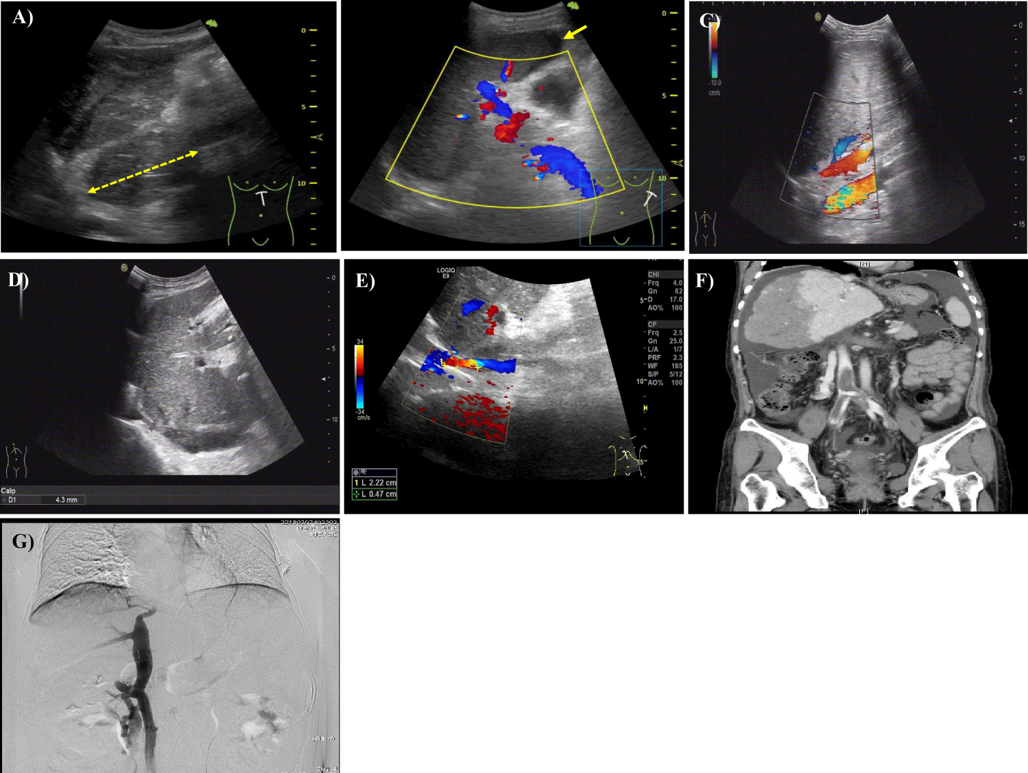
On two-dimensional ultrasound, the hepatic veins were partially or completely absent, and the atresia hepatic veins were strongly echoic in a strip shape, without lumen structure [10]. In case of HV stenosis, the lumen becomes narrow. Color Doppler showed that the outflow tract of hepatic vein was completely obstructed, and there was no blood flow signal at the site of obstruction. Abnormal shaped communicating vessel branches can be seen between the remaining hepatic veins, which can detect bidirectional or reverse blood flow (Figure 1). In case of incomplete obstruction, the blood flow in the obstruction is fine, showing "mosaic" blood flow, and the high speed turbulence spectrum is detected in the lesion.
The manifestation of IVC in BCS patients may be that there is a diaphragm like strong echo in the upper part of the IVC, the echo is uneven, and there may be calcification, and occasionally a sieve shaped sound transmission area on the diaphragm. The manifestations of IVC stenosis and occlusion are similar to those of HV. Ultrasonographic manifestations of HV and IVC in BCS were summed in table 1.
| Table 1: Ultrasonographic manifestations of hepatic vein and inferior vena cava in Budd-Chiari syndrome. | ||
| Gray-Scale Ultrasound | Color And Spectral Doppler | |
| Hepatic vein | ||
| Atresia | The hepatic veins were partially or completely absent, displayed by strongly echoic in a strip shape, without lumen structure | No blood flow signal at the site atresia vein |
| Completely obstruction | Solid echo can be seen in the lumen to completely fill the hepatic vein. The local pipe wall can be thickened with or without thrombus. | No blood flow signal at the site of obstruction |
| Stenosis | Diaphragm like structures can appear in the hepatic vein. Solid echo can be seen in the lumen to partly fill the hepatic vein. | The blood flow in the obstruction is fine, showing "mosaic" blood flow, and the high speed turbulence spectrum is detected at the site of stenosis. |
| Inferior vena cava | ||
| Completely obstruction | Solid echo can be seen in the lumen to completely fill the inferior vena cava. | No blood flow signal at the site of inferior vena cava. |
| Stenosis | Diaphragm like strong echo, uneven echo or calcification, or occasionally a sieve shaped sound transmission area on the diaphragm can be seen in the inferior vena cava. | The blood flow in the obstruction is fine, showing "mosaic" blood flow, and the high speed turbulence spectrum is detected at the site of stenosis. |
Non-specific manifestations include formation of circulation of medial hepatic branches, enlargement of caudate lobe, dilatation of accessory hepatic veins, caudate vein greater than or equal to 3 mm, hepatomegaly, and splenomegaly. Portal vein thrombosis, portal cavernous degeneration, umbilical vein opening, ascites, lower limb vein thrombosis, etc can also be observed.
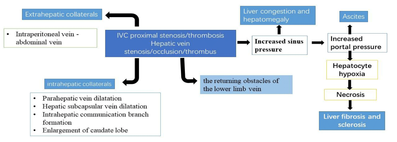
We inferred that the mechanism of these ultrasonic findings may be relevant (Figure 1). The liver of chronic BCS patients is in a state of congestion for a long time due to the continuous obstruction of the HV/IVC outflow tract. Oxygen deficiency can cause the release of oxygen free radicals, causing damage and degeneration of adjacent liver cells, leading to necrosis, fibrosis and sclerosis, and gradually evolving into liver parenchyma lesions, leading to abnormal liver function [11]. BCS is obstructed by HV return. With the increase of internal pressure, HV may dilate, and communicating branches between hepatic veins may form. Most of the blood flow flows from the vein with blocked return to the unobstructed hepatic vein. Color Doppler flow image shows different blood flow directions of several hepatic veins on the same section, and the accessory hepatic veins dilate and form communicating branches with the IVC [12].
Of course, differential diagnosis includes diseases that cross with these specific or nonspecific ultrasonic manifestations, such as liver cirrhosis, hepatitis, liver cancer, right heart failure, constrictive pericarditis, constrictive hypertrophic pleurisy, IVC syndrome. When ultrasound is used for differential diagnosis, it is necessary to know the development history of the patient's condition in detail, and make a comprehensive judgment closely based on clinical manifestations, laboratory tests and other results. For BCS patients, the patient's vascular disease comes first, and liver cirrhosis comes later, so the liver function of patients with acute BCS is generally at a normal level. When pulse hypertension is inconsistent with liver function damage, BCS should be considered. Liver injury occurs first in patients with liver cirrhosis, vascular disease occurs later, and abnormal liver function can occur at the early stage of the disease. The collateral vessels of hepatitis cirrhosis often appear outside the liver, mainly showing the portal systemic circulation pathway, while BCS can show the collateral vessels both inside and outside the liver [12]. No communicating branch and accessory hepatic vein between hepatic veins or HV dilatation were seen in patients with patients of liver cirrhosis.
Special attention should be paid to the ultrasonic examination. The collateral vessels of systemic circulation, the formation of communicating branches between hepatic veins, the expansion of accessory hepatic veins, the stenosis or occlusion of IVC, and the expansion of IVC below the liver segment are all important differential signs between BCS and post hepatitis cirrhosis (Figure 2).
The caudal vein in the third hepatic portal also needs special attention. In healthy people, intrahepatic blood flows back into the inferior vena cava from the second hepatic portal, while a small part of blood flows through the third hepatic portal. The inferior vena cava and these small veins are collectively referred to as accessory hepatic veins in clinical practice. This vein is very small in the ultrasound image of normal people. If one of the caudal veins of accessory hepatic veins can be found in the ultrasound image, with its inner diameter greater than 3 mm [12], BCS should be highly suspected. Some studies have shown that the anterior and posterior diameter of caudate lobe and the diameter of accessory hepatic vein in BCS patients are significantly larger than those in cirrhosis patients. In another study, it was found that the specificity of the combination of specific signs and ‘‘caudate lobe hypertrophy” was 100% [13].
For treatment, drugs based anticoagulation therapy are the mainstay of therapy, although the five-year survival rate after its application is only 30% [14]. Angioplasty is the recommended first-line interventional treatment for patients with BCS. Angioplasty combined with stent placement can improve the restenosis in patients [15]. After failure of angioplasty or stenting a surgical portosystemic shunt or TIPS should be considered. Some serious cases will require liver transplantation [2]. Usually, patients with portal vein thrombosis or portal hypertension may have a poor prognosis [13].
To sum up, color Doppler ultrasound plays a more important role in the diagnosis of patients with BCS and can be popularized in clinical practice. Due to the complexity and variability of intrahepatic vessels and IVC, although there are some significant features, it also requires a certain level of ultrasound doctors.
References
- Ludwig J, Hashimoto E, McGill DB, van Heerden JA. Classification of hepatic venous outflow obstruction: ambiguous terminology of the Budd-Chiari syndrome. Mayo Clin Proc. 1990;65(1):51-5. Epub 1990/01/01. doi: 10.1016/s0025-6196(12)62109-0. PubMed PMID: 2296212.
- Janssen HL, Garcia-Pagan JC, Elias E, Mentha G, Hadengue A, Valla DC, et al. Budd-Chiari syndrome: a review by an expert panel. J Hepatol. 2003;38(3):364-71. Epub 2003/02/15. doi: 10.1016/s0168-8278(02)00434-8. PubMed PMID: 12586305.
- Valla DC. Budd-Chiari syndrome/hepatic venous outflow tract obstruction. Hepatol Int. 2018;12(Suppl 1):168-80. Epub 2017/07/08. doi: 10.1007/s12072-017-9810-5. PubMed PMID: 28685257.
- Wiest I, Teufel A, Ebert MP, Potthoff A, Christen M, Penkala N, et al. [Budd-Chiari syndrome, review and illustration]. Z Gastroenterol. 2022;60(9):1335-45. Epub 2021/11/26. doi: 10.1055/a-1645-2760. PubMed PMID: 34820810.
- Hoffmann T, Voigtlander H, Frohlich E, Debove I, Pauluschke-Frohlich J. Single-center study: evaluation of sonography in Budd-Chiari syndrome. Z Gastroenterol. 2022;60(7):1111-7. Epub 2021/11/16. doi: 10.1055/a-1550-3141. PubMed PMID: 34781388.
- Aydinli M, Bayraktar Y. Budd-Chiari syndrome: etiology, pathogenesis and diagnosis. World J Gastroenterol. 2007;13(19):2693-6. Epub 2007/06/15. doi: 10.3748/wjg.v13.i19.2693. PubMed PMID: 17569137; PubMed Central PMCID: PMCPMC4147117.
- Valla DC, Cazals-Hatem D. Vascular liver diseases on the clinical side: definitions and diagnosis, new concepts. Virchows Arch. 2018;473(1):3-13. Epub 2018/03/25. doi: 10.1007/s00428-018-2331-3. PubMed PMID: 29572606.
- Gupta P, Bansal V, Kumar MP, Sinha SK, Samanta J, Mandavdhare H, et al. Diagnostic accuracy of Doppler ultrasound, CT and MRI in Budd Chiari syndrome: systematic review and meta-analysis. Br J Radiol. 2020;93(1109):20190847. Epub 2020/03/10. doi: 10.1259/bjr.20190847. PubMed PMID: 32150462; PubMed Central PMCID: PMCPMC7217562.
- Bolondi L, Gaiani S, Li Bassi S, Zironi G, Bonino F, Brunetto M, et al. Diagnosis of Budd-Chiari syndrome by pulsed Doppler ultrasound. Gastroenterology. 1991;100(5 Pt 1):1324-31. Epub 1991/05/01. PubMed PMID: 2013376.
- Iliescu L, Toma L, Mercan-Stanciu A, Grumeza M, Dodot M, Isac T, et al. Budd-Chiari syndrome - various etiologies and imagistic findings. A pictorial review. Med Ultrason. 2019;21(3):344-8. Epub 2019/09/03. doi: 10.11152/mu-1921. PubMed PMID: 31476215.
- Goel RM, Johnston EL, Patel KV, Wong T. Budd-Chiari syndrome: investigation, treatment and outcomes. Postgrad Med J. 2015;91(1082):692-7. Epub 2015/10/24. doi: 10.1136/postgradmedj-2015-133402. PubMed PMID: 26494427.
- Bargallo X, Gilabert R, Nicolau C, Garcia-Pagan JC, Ayuso JR, Bru C. Sonography of Budd-Chiari syndrome. AJR Am J Roentgenol. 2006;187(1):W33-41. Epub 2006/06/24. doi: 10.2214/AJR.04.0918. PubMed PMID: 16794137.
- Boozari B, Bahr MJ, Kubicka S, Klempnauer J, Manns MP, Gebel M. Ultrasonography in patients with Budd-Chiari syndrome: diagnostic signs and prognostic implications. J Hepatol. 2008;49(4):572-80. Epub 2008/07/16. doi: 10.1016/j.jhep.2008.04.025. PubMed PMID: 18619699.
- Mancuso A. An update on the management of Budd-Chiari syndrome: the issues of timing and choice of treatment. Eur J Gastroenterol Hepatol. 2015;27(3):200-3. Epub 2015/01/16. doi: 10.1097/MEG.0000000000000282. PubMed PMID: 25590783.
- Wang Q, Li K, He C, Yuan X, Luo B, Qi X, et al. Angioplasty with versus without routine stent placement for Budd-Chiari syndrome: a randomised controlled trial. Lancet Gastroenterol Hepatol. 2019;4(9):686-97. Epub 2019/07/08. doi: 10.1016/s2468-1253(19)30177-3. PubMed PMID: 31279647.
Content Alerts
SignUp to our
Content alerts.
 This work is licensed under a Creative Commons Attribution 4.0 International License.


