Medicine Group . 2023 February 28;4(2):307-309. doi: 10.37871/jbres1676.
Left Parietal Craniectomy in Osteosarcoma with a Collagen Matrix and Cement-Based Repair
Patricia Alejandra Garrido Ruiz1* and Marta Román Garrido2
2Hospital Universitario de Salamanca, P.o de San Vicente, 182, 37007 Salamanca, Primary Care Department, Spain
- Left parietal craniectomy
- Osteosarcoma
- Collagen matrix
Abstract
For routine clinical practice, it is vital prior to surgery to plan the steps of the same as possible complications that may occur and how to resolve them. This case is of special interest for being a large tumor that during its resection, to make it total and facilitate healing, leaves a large defect in the skull shell.
Introduction
On a daily basis, an adhesive collagen matrix coated on its yellow face with fibrinogen and human thrombin is one of the fundamental materials in neurosurgical practice [1-3]. We present a case of osteosarcoma of 8 cm located at the left parietal level. The case is of high interest because of the large size of the lesion and because it is a location that requires craniectomy with exeresis of the lesion and repair of the skull shell [4,5], It is vital to bear in mind the possibility of diagnosis and therapeutic management as well as the planning of the repair of the defect. In addition, in this case, is particularly useful both for facilitating hemostasis and for the correct sealing of the cement cranioplasty with the use of collagen matrix.
Case Report
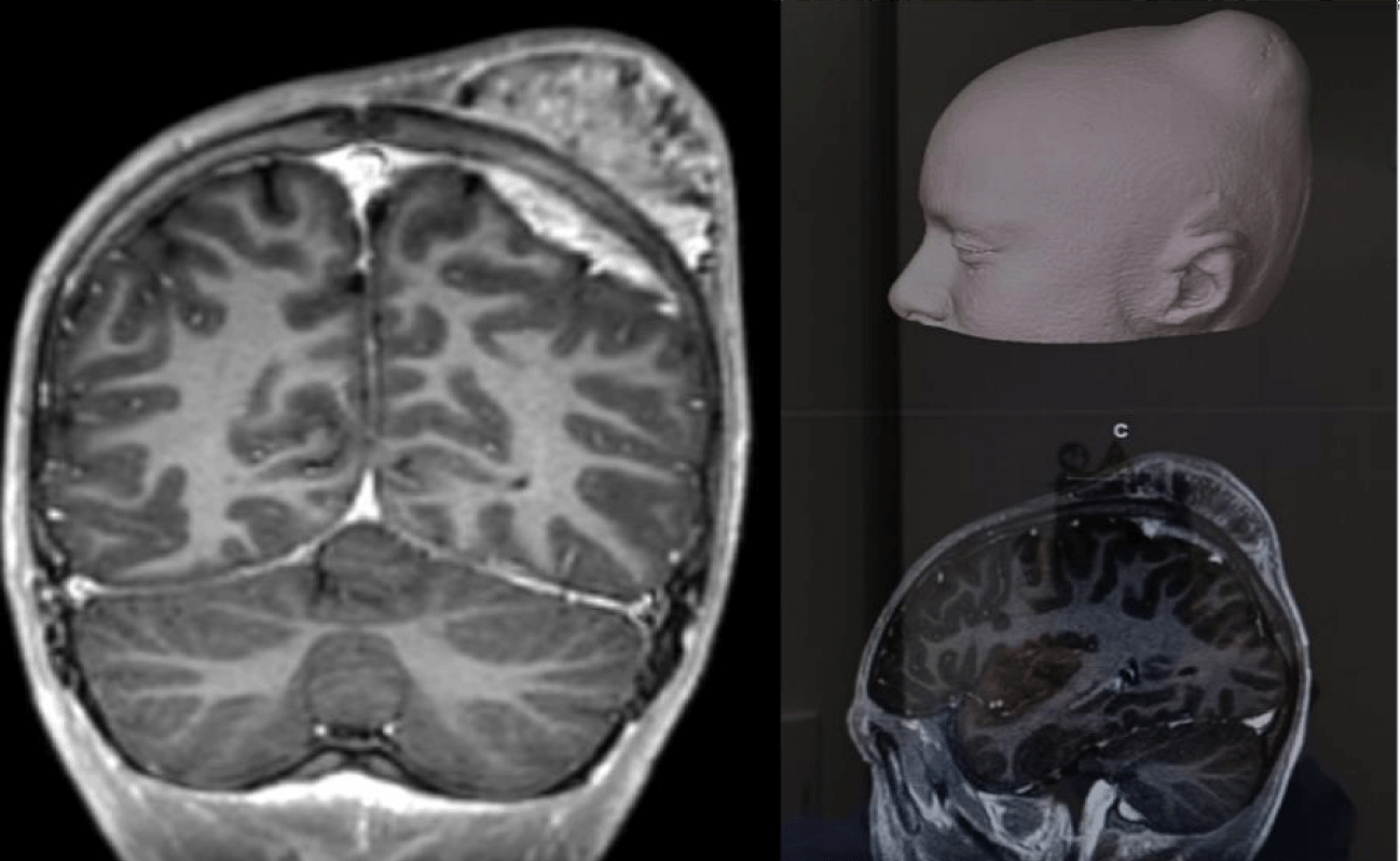
We present the case of a 12-year-old patient who notices a growth of rapid left parietal-occipital growth. No other associated symptomatology. It is performed a brain MRI (Figure 1) that is reported as suggestive of malignancy with a primary bone process in the left parietal bone accompanied by periosteal reaction in external and internal spiculate cortical and a mass of epicranial soft parts of about 8 cm epicranial and towards the intracranial compartment with dural enhancement with differential rhabdomyosarcoma vs. M1 diagnosis of neuroblastoma. No signs of intraspinal extension. A brain CT scan is performed to plan the surgery in which a bone lesion at the left parietal level with an important component of extracranial soft tissue with a bone matrix suggestive of aggressive injury is detected. Bone scan: intensely osteogenic bone lesion with signs of aggressiveness in the left parietal suggestive of Osteosarcoma. No other long-range injuries. After performing a biopsy, he is diagnosed with Osteosarcoma, receiving treatment with chemotherapy protocol SEHOP-10 and Radiotherapy [6-8].
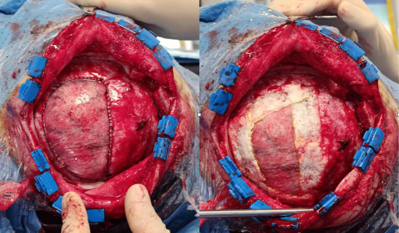
Given the location and malignancy of the lesion, a biparietal incision is made on the tumor. Subperiosteal plane dissection around the lesion. Multiple burr-holes on both sides of the midline. Longitudinal sinus dissection. Block bone resection with the integral lesion. Resection of dura mater in contact with the lesion. Careful hemostasis. Duraplasty with equine pericardium. Above the hemiplasty (Figure 2) on the edges of the dural suture, collagen matrix is placed covering its entire length in a circle. The closure is made by equine pericardium duroplasty, collagen matrix, and hydrogel sealant solution based on polyethylene glycol ester and trilisin amine, with cranioplasty placement with cement fixed with mini-plates and screws.
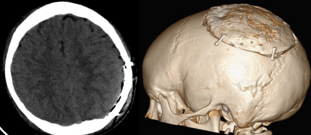
The patient presented a favorable postoperative evolution, with brain CT scans in which no complications were observed (Figure 3), and he remained in the hospital for days after surgery for continued chemotherapy.
Results and Conclusion
We highlight the usefulness of collagen matrix during surgery being fundamental after correct hemostatic and hermetic dural closure sealant prior to the placement of cranioplasty with cement. The case is of high interest because of the large size of the lesion [9,10] and because it is a location that requires craniectomy with exeresis of the lesion and repair of the skull shell. This case report is of interest because it highlights the importance of having into consideration different possibilities when it comes to large defects on the skull after tumor resection.
References
- Youmans. Neurological Surgery. 7th ed. Saunders Company; 2016.
- Rhoton. Rhoton's Cranial Anatomy and Surgical Approaches. Congress of Neurological Surgeons (CNS). Oxford University Press; 2019.
- Greenberg M. Handbook of Neurosurgery. Thieme. 9th ed. 2019.
- Schajowicz F, Sissons HA, Sobin LH. The World Health Organization's histologic classification of bone tumors. A commentary on the second edition. Cancer. 1995 Mar 1;75(5): 1208-14. doi: 10.1002/1097-0142 (19950301) 75: 5<1208::aid-cncr2820750522>3.0.co;2-f. PMID: 7850721.
- Grimer RJ, Taminiau AM, Cannon SR; Surgical Subcommitte of the European Osteosarcoma Intergroup. Surgical outcomes in osteosarcoma. J Bone Joint Surg Br. 2002 Apr;84(3):395-400. doi: 10.1302/0301-620x.84b3.12019. PMID: 12002500.
- Campanacci M. Bone and Soft Tissue Tumors: Clinical Features, Imaging, Pathology and Treatment. 2. Wien, Austria: Springer-Verlag; 1999. p. 464-491.
- Patibandla MR, Uppin SG, Thotakura AK, Panigrahi MK, Challa S. Primary telangiectatic osteosarcoma of occipital bone: a case report and review of literature. Neurol India. 2011 Jan-Feb;59(1):117-9. doi: 10.4103/0028-3886.76891. PMID: 21339678.
- Wolf RE, Enneking WF. The staging and surgery of musculoskeletal neoplasms. Orthop Clin North Am. 1996 Jul;27(3):473-81. PMID: 8649730.
- Huvos AG. Bone Tumors: Diagnosis Treatment and Prognosis. 2. Philadelphia, W.B. Saunders. Co; 1991.
- Huang X, Zhao J, Bai J, Shen H, Zhang B, Deng L, Sun C, Liu Y, Zhang J, Zheng J. Risk and clinicopathological features of osteosarcoma metastasis to the lung: A population-based study. J Bone Oncol. 2019 Mar 7;16:100230. doi: 10.1016/j.jbo.2019.100230. PMID: 30923668; PMCID: PMC6423404.
Content Alerts
SignUp to our
Content alerts.
 This work is licensed under a Creative Commons Attribution 4.0 International License.


