Medicine Group. 2023 December 31;4(12):1755-1763. doi: 10.37871/jbres1862.
Radiological and Chemoteraphy Hints in Ovarian Lymphoma: A Mini Literature Review
Diana Donatello*
Abstract
Introduction: In this mini review we analized some cases of Ovarian Lymphoma (OL) offprint from the previous literature, both primary and secondary.
Discussion: We know that a standard method of staging and follow-up in non-Hodgkin's lymphomas is PET/CT, which, however, due to the limited presence of ovarian lymphomas in the population, is not present in the literature as a primary tool in the diagnosis of this type of neoplasm. The first suspected dignosis is often done with US, CT or MRI, but the gold standard remains the biopsy. As regards for the chemotheraphy those cases poin out the non-toxicity of rituximab and its possible routinary use in the treatment of OL.
Conclusion: US, CT and MRI are the first diagnostic tools in the diagnosis of OL. Nowadays PET/CT is used in the staging and follow-up of OL. The R-CHOP protocol can be utilized in the treatment of OL, reducing the use of readioteraphy. Gynecologists must be aware of this rare ovarian tumor in order to avoid unnecessary radical surgery.
Introduction
The involvement of the ovary in lymphomatous processes is rare, but ovary is the common site in the female genital tract to be involved by the hematological malignancies [1]. The occurrence of lymphomas primarily arising in the ovaries has long been debated since no lymphoid tissue is found in the ovaries [2]. Involvement of the ovary by malignant lymphoma can be primary or secondary and is discovered incidentally during a workup for abdominal or pelvic complaints.
This study highlights some fundamental points, although unfortunately due to the rarity of lymphomatous ovarian manifestations, whether secondary or primary, the numbers available to us to carry out studies are limited and many results are considered statistically insignificant. The points highlighted are:
- The clinical manifestations of ovarian involvement are the same as for other ovarian tumors;
- There is no significant difference in this study in OS (overall survival) and PFS (Progression Free Survival) between primary and secondary lymphoma, therefore the survival estimate is between 59.3% at 5 years for secondary ones and 70.0% at 5 years for primary ones;
- Considering that secondary involvement of the ovary is the result of dissemination of a systemic disease, the OS of secondary lymphoma appears to be better than the overall survival expected for stage III/IV DLBCL lymphoma;
- Burkitt lymphomas respond well to chemotherapy treatment;
- The fact that patients with secondary ovarian involvement respond better than expected may be related to the introduction of rituximab in the treatment, but further studies are necessary to test this hypothesis.
Discussion
Hints on chemoteraphy treatment
Regarding the reported about the use of rituximab in Ovarian Lymphoma (OL), Ahbeddou N, et al. [1], described the case of a 48-year-old patient suffering from ovarian large B-cell lymphoma.
The CT scan at the time of diagnosis revealed that the tumor started from the right ovary, measuring 14 cm in size, and also extended to the greater omentum, the serosa of the fallopian tubes and the mesocolon.
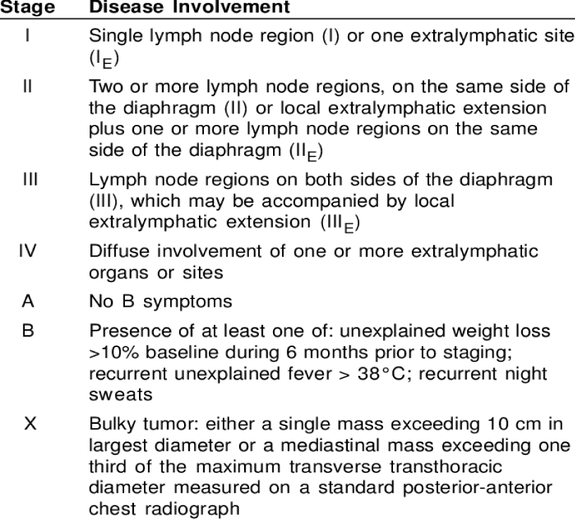
There were no signs of pelvic lymphadenopathy. Bone marrow biopsy was negative. The tumor is assigned an IAEX stage according to the Ann Arbor classification and a stage II, according to the FIGO classification. The IPI, the International Prognostic Index, was low.
The chemotherapy treatment in this case consists of the R-CHOP regimen (rituximab 375 mg/m2, cyclophosphamide 750mg/m2, doxorubicin 50mg/m2, vincristine 1.4mg/m2 and prednisolone 100mg every week). The CT performed after six cycles of chemotherapy shows a complete remission. The patient is therefore asymptomatic, without evidence of disease, and with subsequent regular follow-up.
We remind you of some useful prognostic factors in ovarian lymphomas:
- The clinical stage;
- The mode of onset;
- The histological type;
- The IPI index.
Regarding the stage, some previous studiesmhighlighted that the Ann Arbor staging system is inadequate for extranodal lymphomas. The FIGO system seems to be more adequate and gives greater benefits and used in combination with the Ann Arbor system, or another more precise measurement system would be necessary.
Furthermore, we know that there is no standard protocol for the treatment of primary or secondary ovarian non-Hodgkin lymphomas [2].
The best common opinion seems to be to use chemotherapy in accordance with the histological type.
Niitsu N, et al. [3] reported already in 2002 a case of 54-year-old Japanese female with abnormal genital bleeding. Pelvic CT and MRI showed a right ovarian tumor with a diameter of 7 cm and an irregular border. With the diagnosis of a right ovarian tumor, the patient underwent a simple hysterectomy and bilateral salpingo-oophorectomy. Microscopic examination of the right ovarian tumor revealed vaguely nodular growth of small lymphoid cells. They were CD10+, Bcl-2+, Cyclin D1- and CD21-, although CD21+ follicular dendritic cell clusters were present as a background component in each vague nodule. A conventional cytogenetic analysis revealed t(14;18)(q32;q21), and somatic hypermutation of the variable region of the immunoglobulin heavy chain Gene (IgH) was confirmed by direct sequencing of subcloned DNA samples derived from PCR amplicons. These findings led us to characterize the lesion as follicular lymphoma, grade 1. The patient was free of detectable disease 9 months after initiation of post-surgical chemotherapy treated using different combinations of chemotherapy compared to R-CHOP: cyclophosphamide, vincristine, bleiomycin, etoposide, doxorubicin and prednisolone.
Ahbeddou's case highlights the non-toxicity of rituximab and its possible routine use in the treatment of ovarian lymphomas. As the author points out, gynecologists must be aware of this rare ovarian manifestation in order to avoid unnecessary radical surgery.
Signorelli M, et al. [2] reviewed from 1984 to 2003, the treatments in there institution of 19 patients with genital lymphoma. Nine women presented with cervical, 3 with vaginal, 1 with cervical-vaginal, 2 with vulvar and 4 with ovarian lymphoma. Seven were staged IE, nine IIE, one IIIE and two IVE. As a whole, chemotherapy was used in 18/19 cases: chemotherapy was proposed as first line treatment in 12 cases, while surgery in 7 (followed by chemotherapy in 6 cases). Primary chemotherapy alone obtained a Complete Response (CR) in 9/12 patients; Pathological Complete Response (pCR) was confirmed in 3 operated patients out of 9. Partial Response (PR) was observed in 3, requiring radiotherapy. Chemotherapy obtained CR after incomplete surgical debulking in 3 out of 4 cases. Two patients relapsed in the group treated with chemotherapy alone. Both have been salvaged by further chemotherapy. Only one patient deceased due to her tumor after surgery and chemotherapy.The use of exclusive chemotherapy obtained promising results not only as regards survival rates but also for reducing the need of radiotherapy. A conservative management based on exclusive chemotherapy in primary genital lymphoma stages I-II may be attempted in selected patients desiring pregnancy.
PET in staging and follow-up control after chemotherapy treatment
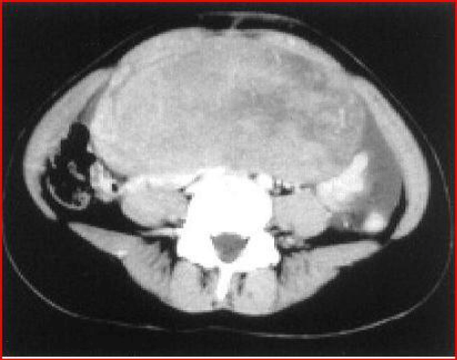
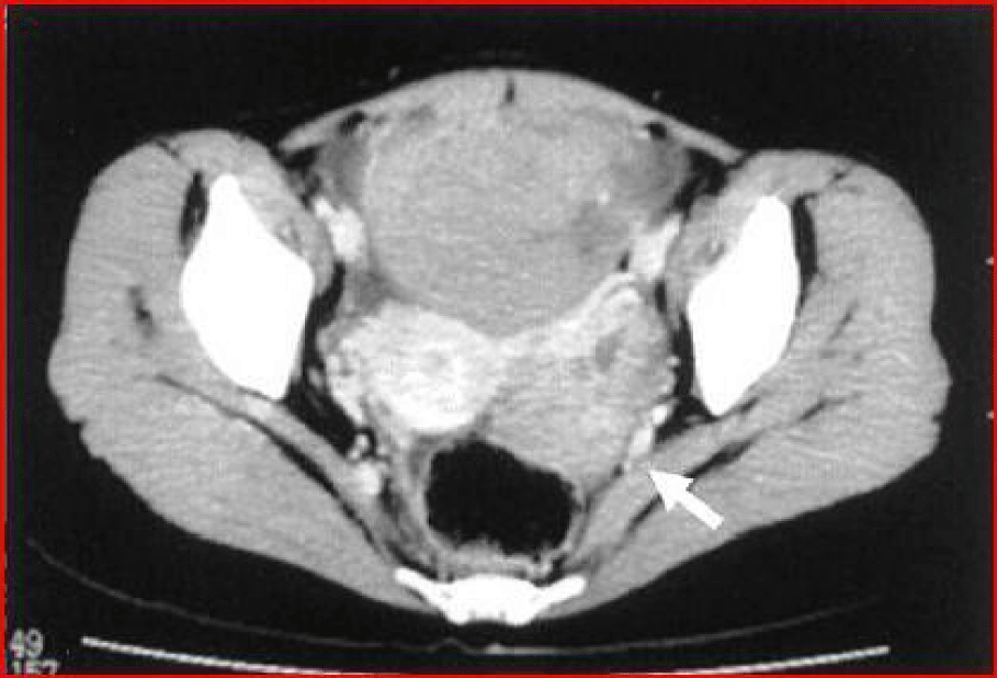
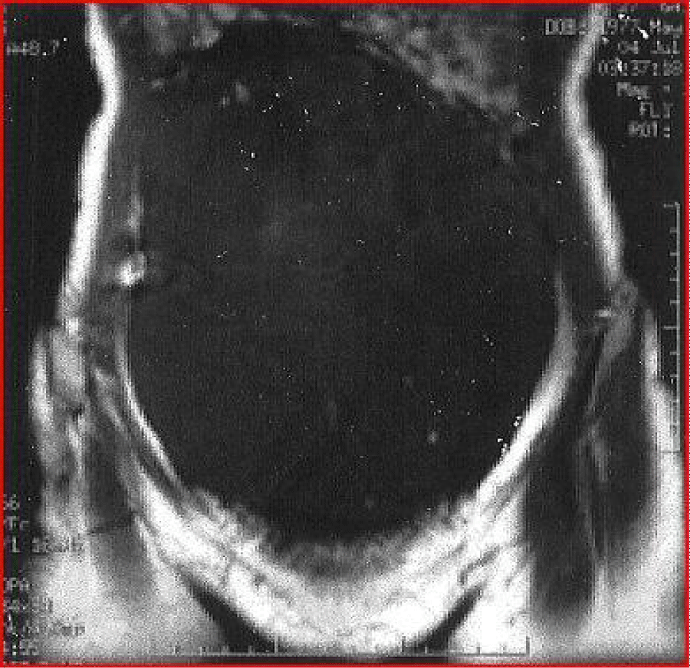
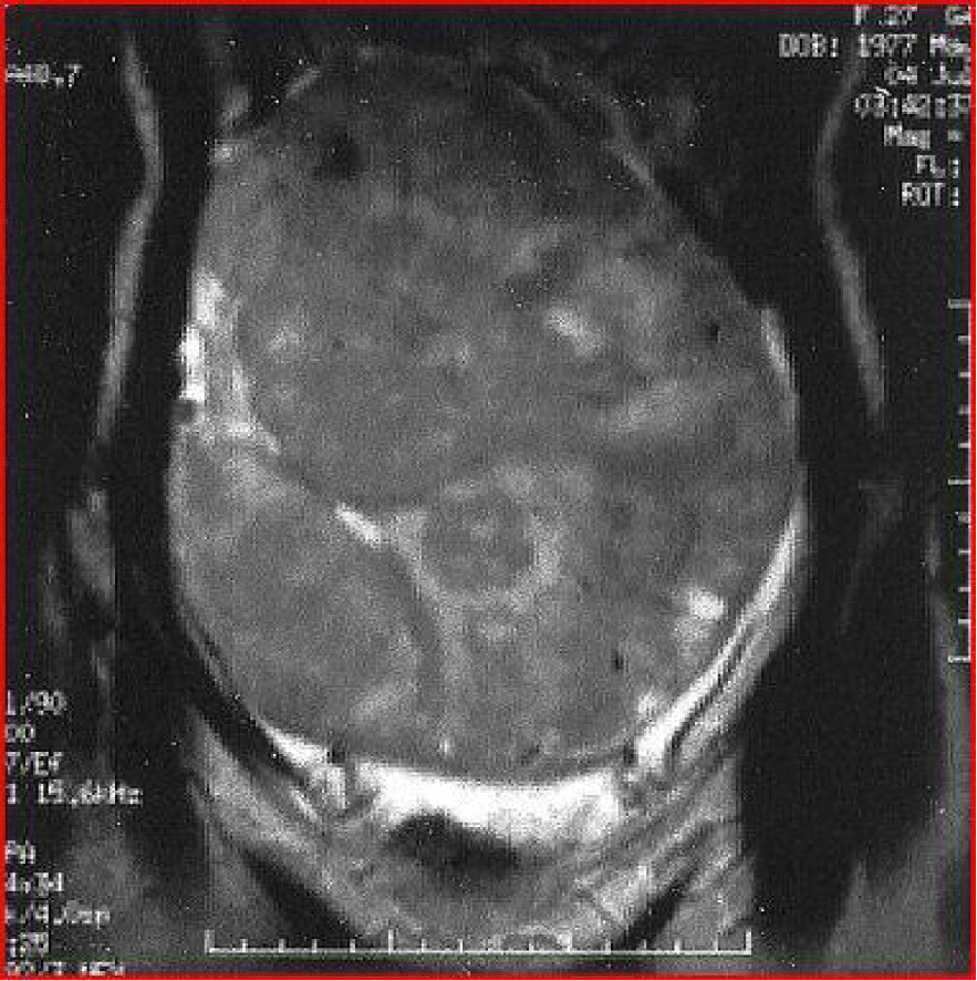
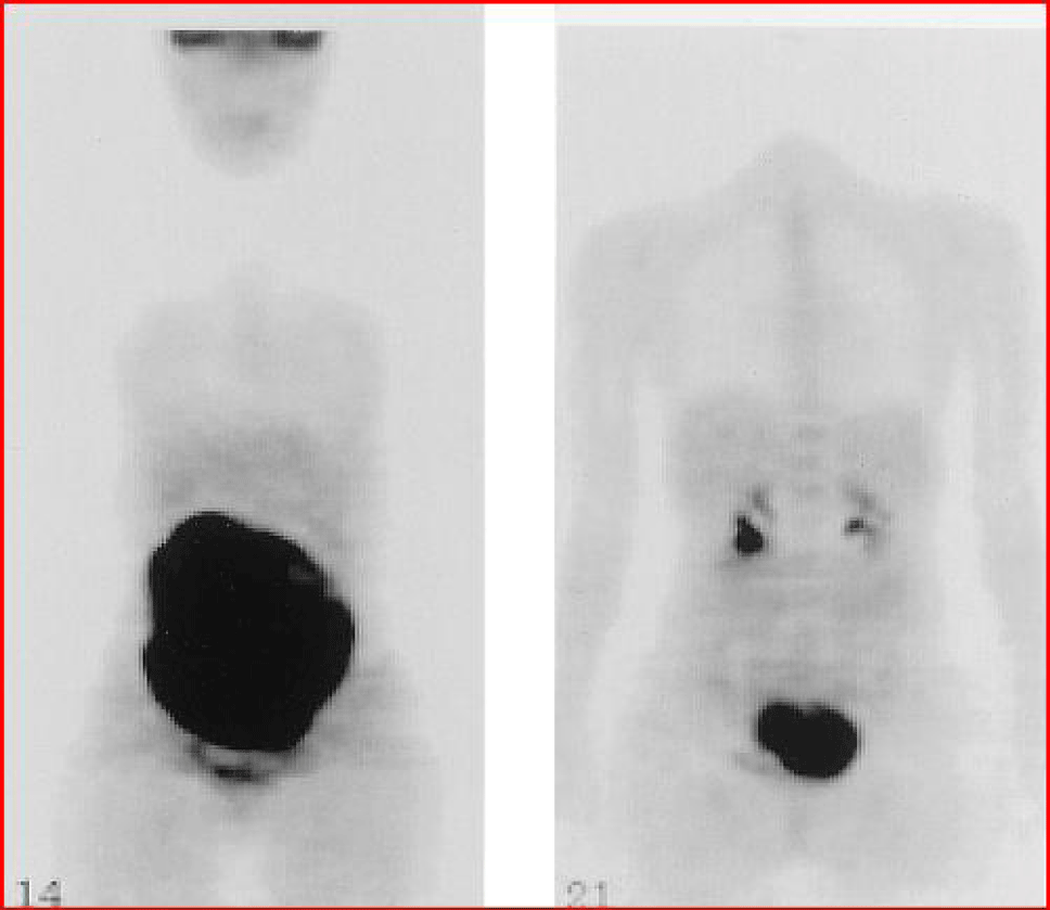
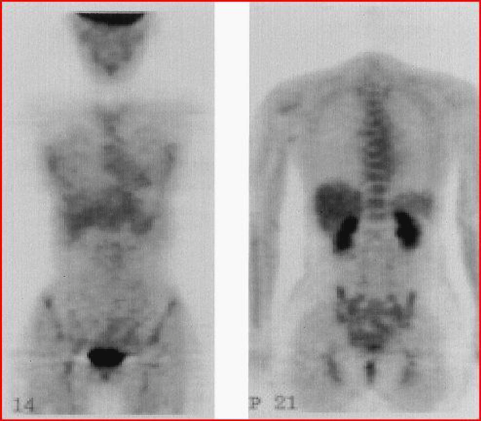
We observed the radiological images obtained by Daisuke Komoto D, et al. [4] that point out in detail the presence of a primary ovarian B-cell lymphoma (Figures 1-4). In their case, during exploratory laparoscopy the left uterus and ovary are preserved given the young age of the patient. Once the histochemical analysis has been carried out and it has been ascertained that it was a stage IV diffuse B-cell lymphoma with an IPI of three (intermediate-high risk), the patient is subjected to three cycles of R-CHOP (rituximab, cyclophosphamide, doxorubicin, vincristine and prednisolone). CT and 18F-FDG PET three months after treatment reveal no signs of involvement. As we well know, a standard method of staging and follow-up in non-Hodgkin's lymphomas is PET/CT, which, however, obviously due to the limited presence of ovarian lymphomas in the population, is not present in the literature as a primary means in the diagnosis of this type of neoplasm. Komoto in his 2006 article opens the doors to the use of 18F-FDG PET in the research and identification of ovarian lymphoma, demonstrating a strong up-take by the ovarian mass with a max SUV equal to 12.5 (Figure 5). As we observe in the latest images shown, PET shows that after chemotherapy treatment the left ovarian mass has significantly reduced (Figure 6). It is therefore useful to report this single case as a reference point for monitoring the therapeutic response in patients affected by ovarian lymphoma.
Conclusion
The R-CHOP protocol can be used in the treatment of OL and the PET/CT is a useful tools in the diagnosis and follow-up of OL.
rom 1984 to 2003, the treatments in our institution of 19 patients with genital lymphoma were reviewed. Nine women presented with cervical, 3 with vaginal, 1 with cervical-vaginal, 2 with vulvar and 4 with ovarian lymphoma. Seven were staged IE, nine IIE, one IIIE and two IVE. As a whole, chemotherapy was used in 18/19 cases: chemotherapy was proposed as first line treatment in 12 cases, while surgery in 7 (followed by chemotherapy in 6 cases).
The use of exclusive chemotherapy obtained promising results not only as regards survival rates but also for reducing the need of radiotherapy. A conservative management based on exclusive chemotherapy in primary genital lymphoma stages I-II may be attempted in selected patients desiring pregnancy.
Finally gynecologists must be aware of this rare ovarian tumor in order to avoid unnecessary radical surgery.
The R-CHOP protocol appears to be superior to CHOP alone in terms of response and relapse rate in the case of secondary involvement.
References
- Ahbeddou N, Fetohi M, Khanoussi BE, Errihani H. Rituximab CHOP for successful management of diffuse large B-cell lymphoma of the ovary. Arch Gynecol Obstet. 2011 May;283(5):1173-4. doi: 10.1007/s00404-010-1701-0. Epub 2010 Oct 19. PMID: 20957380.
- Signorelli M, Maneo A, Cammarota S, Isimbaldi G, Garcia Parra R, Perego P, Maria Pogliani E, Mangioni C. Conservative management in primary genital lymphomas: the role of chemotherapy. Gynecol Oncol. 2007 Feb;104(2):416-21. doi: 10.1016/j.ygyno.2006.08.024. Epub 2006 Oct 16. PMID: 17049970.
- Niitsu N, Nakamine H, Hayama M, Unno Y, Nakamura S, Horie R, Iwabuchi K, Nakamura N, Miura I, Higashihara M. Ovarian follicular lymphoma: a case report and review of the literature. Ann Hematol. 2002 Nov;81(11):654-8. doi: 10.1007/s00277-002-0545-5. Epub 2002 Oct 12. PMID: 12454705.
- Komoto D, Nishiyama Y, Yamamoto Y, Monden T, Sasakawa Y, Toyama Y, Satoh K, Ohno M, Kanenishi K, Ohkawa M. A case of non-hodgkin's lymphoma of the ovary: usefulness of 18F-FDG PET for staging and assessment of the therapeutic response. Ann Nucl Med. 2006 Feb;20(2):157-60. doi: 10.1007/BF02985629. PMID: 16615426.
Content Alerts
SignUp to our
Content alerts.
 This work is licensed under a Creative Commons Attribution 4.0 International License.


