> Medicine. 2020 June 04;1(1):008-010. doi: 10.37871/jels1113.
Dermoid Cyst of the Frontal Bone: A Case Report
Beghdad Mohamed*, Ballage Amine, Mkhatri Amine, Chaouki Anas, Oukessou Youssef, Rouadi Sami, Abada Reda, Roubal Mohamed and Mahtar Mohamed
- Dermoid cyst
- Frontal bone
- Frontal sinus
Introduction: Dermoid cysts are benign tumors that originate from aberrant primordial tissues. About 7% of all dermoid cysts are located in the head and neck region. We present here the case of a periorbital dermoid cyst in a 34 years old patient, involving the frontal bone.
Case Report: We report the case of a 34 years old female patient, who has a history of a cystic formation of the superior inner angle of the right orbit, for which she received surgery 3 years prior. Clinical examination found a swelling located in the superior inner angle of the right eye, of hard consistency, painless, without local inflammatory signs, measuringapproximatively 2 cm. A CT-scan was realized, showing images of an encapsulated and limited supraorbital lesion, developed on both sides of the frontal bone, with an external component. Histopathology examination revealed the presence of keratinized squamous cells, confirming the diagnosis of a dermoid cyst.
Conclusion: Frontal dermoid cysts are uncommon but need to be considered as a differential diagnosis of any mass in orbital region. CT can help to suggest the diagnosis. Its surgical excision must be complete to avoid recurrence.
Dermoid cysts are benign tumors that originate from aberrant primordial tissues [1]. They are formed by dermal and epithelial cells, which result from the entrapment of ectodermal tissue during embryologic development [2].
About 7% of all dermoid cysts are located in the head and neck region. Common locations include the periorbital, nasal, scalp and postauricular regions [3]. We present here the case of a periorbitaldermoidcyst in a 34 yearsold patient, involving the frontal bone.
We report the case of a 34 years old female patient, who has a history of a cystic formation of the superior inner angle of the right orbit, for which she received surgery 3 years prior. The exact nature of this mass is unknown, as the patient was treated at a different hospital, and did not have any document relevant to the histology of the lesion.
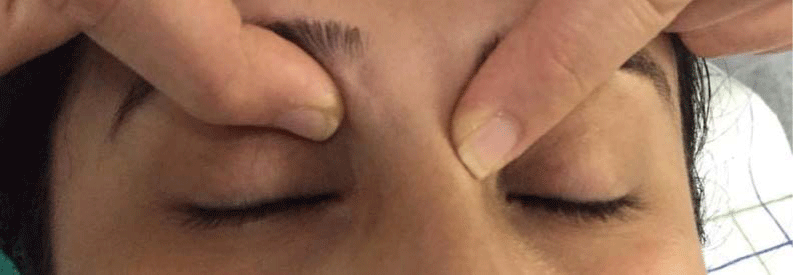
This patient presented to our department for a recurrence of the mass 3 months after the surgery. Clinical examination found a swelling located in the superior inner angle of the right eye, of hard consistency, painless, without local inflammatory signs, measuring approximatively 2 cm (Figure 1).
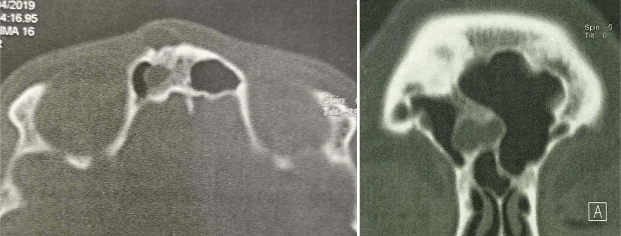
A CT-scan was realized, showing images of an encapsulated and limited supraorbital lesion, developed on both sides of the frontal bone, with an external component measuring 12 x 7 mm, and an internal component in the frontal sinus, measuring 14 x 7 mm (Figure 2).
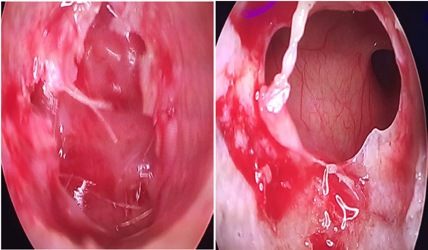
The patient received surgical treatment; we first performed a periorbital incision, and excised the external component of the cyst (Figure 3); then, the frontal sinus was accessed through the nasal cavity, allowing for the removal of the internal component, which contained keratin-like material.
Histopathology examination revealed the presence of keratinized squamous cells, confirming the diagnosis of a dermoid cyst.
Dermoid cysts within the ocular region are thought to arise from sequestered benign ectodermal tissue, often at suture lines. The most common involved suture lines are the zygomatico-frontal suture in 2/3 and the fronto-ethmoidal suture in 1/3 of patients [4,5] .Orbital dermoids frequently have an intracranial extension. Extension into the frontal sinus has also been reported in the literature [6]. Dermoid cysts must be considered as part of the differential diagnosis of any mass in the orbital region [7,8]. In general, these lesions present in childhood. When they occur deep within the orbit, they may escape diagnosis until adulthood when they present with ophthalmologic symptoms as well as erosion of nearby bony tissues [9].
Given the embryologic origin of dermoid cysts, it is essential for those presenting as a midline mass of the skull or scalp to be comprehensively evaluated preoperatively, including CT and/or MRI to assess for intracranial involvement [2].
Histologically, they have a lining of squamous epithelium with dermal elements such as hair follicles, sebaceous, and sweat glands. Within the cyst, keratin, hair, smooth muscle, and lipid debris can be found.
Surgical excision of dermoid cysts is a common intervention. The surgeon must try to maintain the cyst wall intact during the surgical excision, since the inflammatory process may occur if its contents remain within the orbit [10]. Total removal of the entire epithelial lining and the stalk or tract attached to the wound base is necessary to ensure no recurrence [1].
Histologically, they have a lining of squamous epithelium with dermal elements such as hair follicles, sebaceous, and sweat glands. Within the cyst, keratin, hair, smooth muscle, and lipid debris can be found [9].
Frontal dermoid cysts are uncommon but need to be considered as a differential diagnosis of any mass in orbital region. CT can help to suggest the diagnosis. Its surgical excision must be complete to avoid recurrence.
- Sadeghi Tari A, Eshraghi B, Torabi HR. Dermoid cyst of the frontal bone: A case report. IRJO. 2011; 23: 57-59.
- Wood J, Couture D, David LR. Midline dermoid cyst resulting in frontal bone erosion. J Craniofac Surg. 2012; 23: 131-134. https://bit.ly/2ABk87t
- Splendiani A, Bruno F, Mariani S, La Marra A, Capretti I, Di Cesare E, et al. A rare localization of pure dermoid cyst in the frontal bone. Neuroradiol J avr. 2016; 29: 130-133. https://pubmed.ncbi.nlm.nih.gov/26915898/
- Abou-Rayyah Y, Rose GE, Konrad H, Chawla SJ, Moseley IF. Clinical, radiological and pathological examination of periocular dermoid cysts: evidence of inflammation from an early age. Eye. 2002; 16: 507-12. https://pubmed.ncbi.nlm.nih.gov/12194059/
- Sherman RP, Rootman J, Lapointe JS. Orbital dermoids: clinical presentation and management. Br J Ophthalmol. 1984; 68: 642-652. https://pubmed.ncbi.nlm.nih.gov/6466593/
- Pryor SG, Lewis JE, Weaver AL, Orvidas LJ. Pediatric dermoid cysts of the head and neck. Otolaryngol Neck Surg. Juin. 2005; 132: 938-942. https://bit.ly/2U5CCEi
- Ohtsuka K, Hashimoto M, Suzuki Y. A review of 244 orbital tumors in Japanese patients during a 21-year period: origins and locations. Jpn J Ophthalmol. Fevr. 2005; 49: 49-55. https://pubmed.ncbi.nlm.nih.gov/15692775/
- Bonavolonta G, Strianese D, Grassi P, Comune C, Tranfa F, Uccello G, et al. An Analysis of 2,480 Space-Occupying Lesions of the Orbit from 1976 to 2011. Ophthal Plast Reconstr Surg. 2013; 29: 79-86. https://bit.ly/36V8Xm9
- Pham N, Dublin A, Strong E. Dermoid Cyst of the Orbit and Frontal Sinus: A Case Report. Skull Base. Juill. 2010; 20: 275-278. https://pubmed.ncbi.nlm.nih.gov/21311621/
- Correa Perez ME, Sanchez-Tocino H, Blanco Mateos G. Dermoid cyst in childhood, diagnosed as ptosis. Arch Soc Esp Oftalmol Engl Ed. 2010; 85: 215-217. https://pubmed.ncbi.nlm.nih.gov/21074097/
Content Alerts
SignUp to our
Content alerts.
 This work is licensed under a Creative Commons Attribution 4.0 International License.


 Save to Mendeley