Medicine Group . 2022 January 26;3(1):082-084. doi: 10.37871/jbres1403.
Right Atrial Myxoma in Patients with Metastatic Breast Cancer: Multidisciplinary Approach and Surgical Timing
Vite Giuseppe Alessio, Ceresa Fabrizio*, Mammana Liborio Francesco, De Donno Filomena Bruna, Rubino Antonino Salvatore and Patane Francesco
Abstract
Primary cardiac tumours are very rare. We describe the case of a 69-year-old female suffered from metastatic breast cancer and intestinal polyposis. During periodical follow-up examinations, total body Computed Tomography showed a right atrial mass with heterogeneous contrast enhancement, located near the orifice of inferior vena cava. The trans-thoracic and trans-esophageal echocardiogram confirmed the diagnosis. Thrombus or cardiac metastatic localization was the two principal hypotheses even if the CT scan suggested that was a myxoma. Surgical resection was performed expecially for the high risk of the mass' embolization. The hystological examination confirmed the CT scans suspiction of cardiac myxoma. The patient was discharged from hospital without any complications. After 6 months, she was fine.
Introduction
The primary cardiac tumours are very rare [1]. Cardiac myxoma is the most common of them and can lead to several complications as embolization, intra cardiac obstructions, conduction disturbances and lethal valve obstructions [2-5]. Cardiac myxoma is usually located in the left atrium and when it arises from the right chambers, lipoma [6] or the less common malignant primary cardiac tumours should be ruled out.
Case Report
A 69-year-old female was referred to our centre for the right atrial mass. Her past medical history revealed previous laparotomic cholecystectomy, hypothyroidism due to multinodular goiter, Parkinson's disease and osteoporosis. In the 2014, she was operated on right quadrantectomy for breast cancer with subsequent chemotherapy and radiotherapy treatments. During follow-up period, she underwent a periodical total body Computed Tomography (CT) that showed a good therapy response without the onset of metastatic lesions. In the 2018, she suffered from a severe anemia with faecal occult blood and, so, she underwent gastroscopy and colonoscopy that showed a diffuse gastrointestinal polyposis. A multi-lobed polyps located in the right colon was endoscopically resected and its histological examination confirmed the diagnosis of adenomatous intestinal polyp. The next year the abdominal CT showed the onset of hepatic nodular formations at the VIII and VII segments that were dealt with a laparoscopic biopsy, confirming the suspicion of carcinomatous metastasis. The neo-adjuvant chemotherapy was immediately started and the hepatic secondary lesions remained stable for a long time. After beginning this therapy, an echocardiogram was yearly performed.
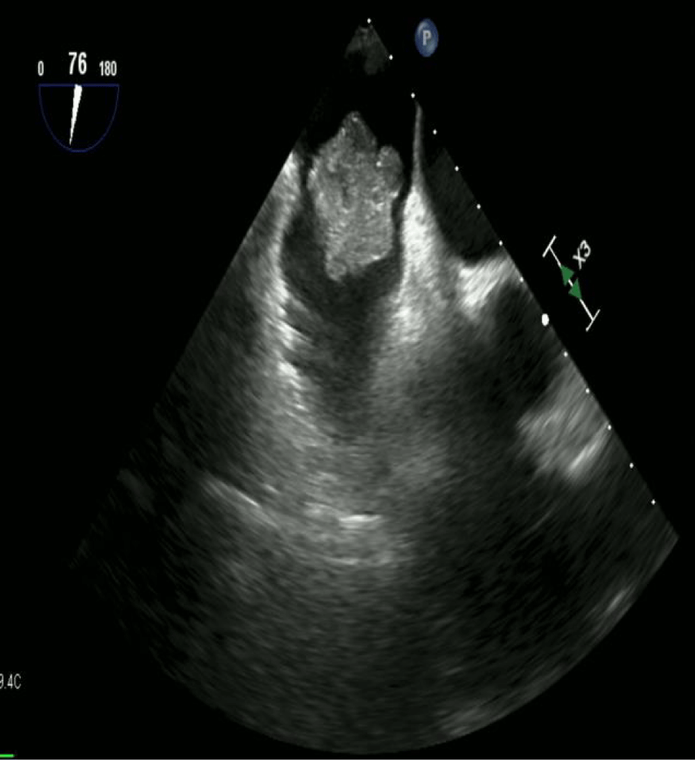
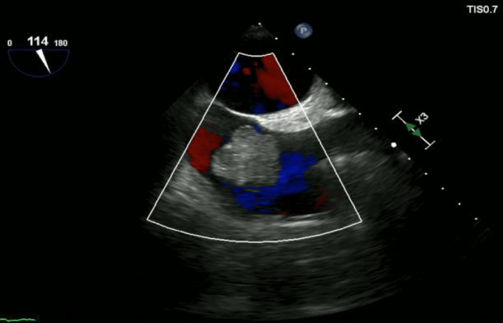
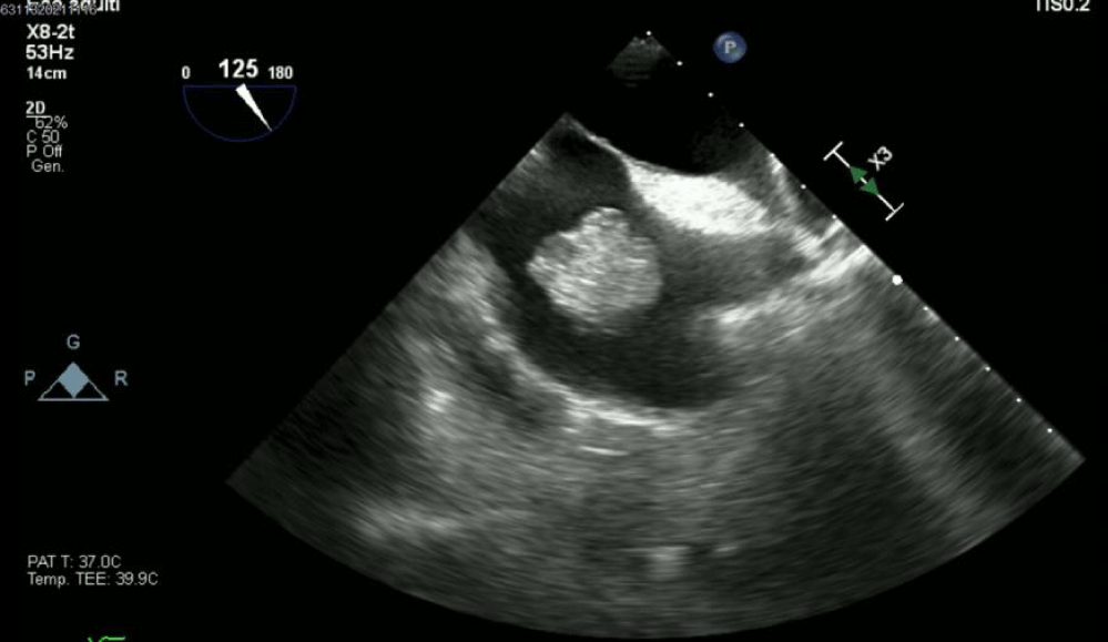
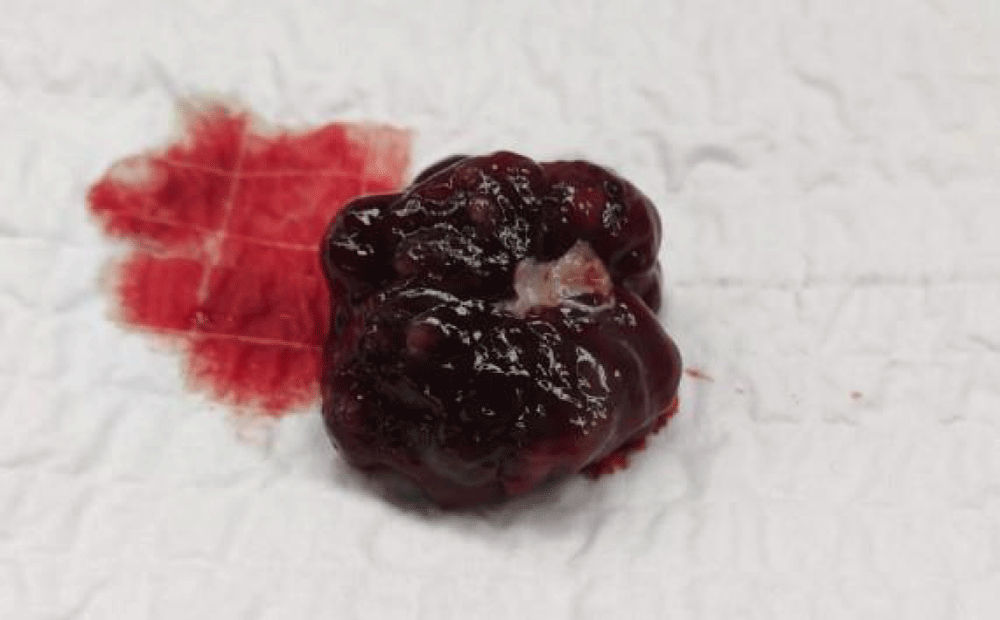
In the 2021, the annual total body CT showed a mass about of 2.5 cm near the junction between inferior vena cava and the right atrium. The mass appeared as intracavitary filling defects with heterogeneous contrast enhancement. The neoplastic history of the patient suggested a tumoral nature of the mass, but we couldn't exclude that was a thrombus. A trans-thoracic (Figures 1,2) and a trans-esophageal (Figure 3) echocardiograms showed a very mobile pedicle right atrial mass about of 3 cm of diameter and its relationship with the Eustachian valve, tricuspid valve and inferior vena cava's orifice. We would have liked to perform a cardiac Magnetic Resonance (MR), but the patient refused because she suffered from claustrophobia. Surgical indication was given to remove the right intra-atrial mass; especially for the high risk of emobolization. A median sternotomy was performed. Cardiopulmonary bypass was initiated with distal ascending aorta cannulation and bicaval venous cannulation. The tumor appeared to arise from the fossa ovalis near to the orifice of the inferior vena cava. The mass and the part of atrial septum infiltrated were completely removed. We preferred to close the surgical defect with a direct running suture as the size of the septal aneurysm allowed for it. The histological examination confirmed the diagnosis of cardiac myxoma (Figure 4). The patients were discharged from hospital without any problems after one week.
Discussion
The primary cardiac tumour are very rare [1], ranging from 0.001 to 0.28% of all tumours [6]. The cardiac myxomas are approximately 80% of all cardiac tumours, occurring mainly in the 3rd–6th decade of life [1-5,6]. The transthoracic echocardiography remains an important tool to detect atrial myxoma, having got sensitivity about of 90%, that becomes higher in the case of trans-esophageal examination [5]. The atrial myxomatous formations usually occur in the left atrium and in very few cases in the right cardiac chambers. We want to underline certain features of the management of our case. First, the role of CT scans to identify the nature of the cardiac tumours and their relationship with the near structures, allowing planning the best surgical approach. Second, we must keep in mind that heart can be involved as metastatic localization: the growth of the tumour is usually slow and it can often remain asymptomatic for long time. In particular way, the history of our patient, suffered from metastatic breast cancer and intestinal polyposis, could suggest a cardiac metastatic localization to explain the genesis of right atrial mass. Another hypothesis could be a para-neoplastic thrombus. Despite an echocardiogram's high sensitivity to detect the intra-cardiac masses, contrast-enhanced cardiac CT seems to be more able to discriminating between a thrombus and cardiac tumours, even if probably the cardiac MR remains the gold standard for this disease.
Conclusion
The histological examination confirmed the diagnosis of myxoma as it had already been suspected with cardiac CT. In conclusion, in case of cardiac mass when it is impossible to perform a cardiac MR, a contrast-enhanced CT can give a lot of information useful to plan the surgical strategy.
References
- Centofanti P, Di Rosa E, Deorsola L, Dato GM, Patanè F, La Torre M, Barbato L, Verzini A, Fortunato G, di Summa M. Primary cardiac tumors: Early and late results of surgical treatment in 91 patients. Ann Thorac Surg. 1999 Oct;68(4):1236-1241. doi: 10.1016/s0003-4975(99)00700-6. PMID: 10543485.
- Jelic J, Milicić D, Alfirević I, Anić D, Baudoin Z, Bulat C, Corić V, Dadić D, Husar J, Ivanćan V, Korda Z, Letica D, Predrijevac M, Ugljen R, Vućemilo I. Cardiac myxoma: Diagnostic approach, surgical treatment and follow-up. A twenty years experience. J Cardiovasc Surg (Torino). 1996 Dec;37(6 Suppl 1):113-117. PMID: 10064362.
- Vicari RM, Polanco E, Schechtmann N, Santiago JO, Shaurya K, Halstead M, Marszal D, Grosskreutz T, Thareja S. Atrial myxoma presenting with orthostatic hypotension in an 84-year-old Hispanic man: A case report. J Med Case Rep. 2009 Dec 14;3:9328. doi: 10.1186/1752-1947-3-9328. PMID: 20062757; PMCID: PMC2803851.
- Reynen K. Cardiac myxomas. N Engl J Med. 1995 Dec 14;333(24):1610-1617. doi: 10.1056/NEJM199512143332407. PMID: 7477198.
- Mügge A, Daniel WG, Haverich A, Lichtlen PR. Diagnosis of noninfective cardiac mass lesions by two-dimensional echocardiography. Comparison of the transthoracic and transesophageal approaches. Circulation. 1991 Jan;83(1):70-78. doi: 10.1161/01.cir.83.1.70. PMID: 1984900.
- Ceresa F, Calarco G, Franzì E, Patanè F. Right atrial lipoma in patient with Cowden syndrome. Interact Cardiovasc Thorac Surg. 2010 Dec;11(6):803-804. doi: 10.1510/icvts.2010.245001. Epub 2010 Sep 19. PMID: 20852328.
Content Alerts
SignUp to our
Content alerts.
 This work is licensed under a Creative Commons Attribution 4.0 International License.


