Medicine Group . 2022 November 17;3(11):1321-1325. doi: 10.37871/jbres1600.
Minimally Invasive Surgical Approach to Decompress a Multi-Loculated Abscess in the Thoracic Spine: A Case Report
Andre Evaristo Marcondes Cesar1-4*, Leonardo Mousinho4,5, Ricardo Rodriguez Ferreira6,7, Lucas Ponich Clementino6, Wilker Herkson de Almeida Oliveira8, Tiago Ernesto Fabris Cezar9,10, Blanca Elena Guerrero Daboin11 and Luciano Miller Reis Rodrigues2
2Group of Spine Surgery of School of Medicine Centro Universitario ABC, Santo Andre, SP, Brazil
3Department of Spine Surgery at Associação de Assist0ência à Criança Deficiente (AACD), São Paulo, SP, Brazil
4Centro de Ortopedia, Traumatologia e Coluna (COTC), Sao Paulo, SP, Brazil
5Hospital Estadual Mario Covas, R. Dr. Henrique Calderazzo, Santo Andre, SP, Brazil
6Neurophysiology Clinic, São Paulo, SP, Brazil.
7Institute of Orthopedics and Traumatology, Universidade de São Paulo, SP, Brazil.
8Spine Surgery Fellowship - School of Medicine, Centro Universitario A.B.C., Santo Andre, SP, Brazil
9Department of Thoracic Surgery- Universidade Federal de Campinas, SP, Brazil
10Department of Thoracic Oncology- Universidade de São Paulo, SP, Brazil
11Laboratory and writing center of Postgraduate Studies, School of Medicine Centro Universitario A.B.C., Santo Andre, SP, Brazil
- Tuberculosis
- Extrapulmonary tuberculosis
- Pott disease
- Video-assisted surgery
Abstract
Tuberculosis (TB) remains one of the major global causes of death; Mycobacterium Tuberculosis causes it. Approximately 10% of patients with TB may develop extrapulmonary bone tuberculosis, with the spine being the most affected, representing 1 to 2% of all TB cases. Pott disease is a form of extrapulmonary TB, and recently it has shown a significant resurgence in developed nations possibly impacted by global migration. Pott disease is often misdiagnosed or sometimes diagnosed during its late stages, which can be associated with devastating consequences to the patient. Brazil is among the top 20 countries with a high burden of tuberculosis; however, the published evidence is still limited, particularly during the last two years of the COVID-19 pandemic. Under this context, we present a clinical case of a 43 years old female patient with Pott disease with a minimally invasive surgical approach to decompress a multi-loculated abscess in the thoracic spine.
Introduction
Tuberculosis (TB) remains one of the major global causes of death. In 2020, this infectious disease killed almost ten million people worldwide, most of them in their productive years (WHO-a) [1]. More than 95% of deaths and cases occur in developing countries.
Predominantly TB affects the lungs, but during its evolution from the latent phase to the active stage, it can affect other body organs, defined as Extrapulmonary Tuberculosis (EPTB) [2].
In 2017, 16% of the 6.3 million TB cases identified by WHO were extrapulmonary TB cases [3]. Spinal TB, or Pott disease, a form of EPTB, has recently shown a marked revival in developed nations, perhaps influenced by transnational migration [4].
In Brazil, the evidence published about EPTB, specifically Pott disease, is still limited. It could be due to the challenges of establishing a diagnosis in the early stages of the illness. Hence, this paper aims to present a clinical case of Pott disease with a minimally invasive surgical approach to decompress a multi-loculated abscess in the thoracic spine.
Patient information
The patient, 43 years old, female in good health and without comorbidities, reported excruciating and progressive pain complaints in the dorsal spine region over the last 06 months, without signs of trauma history, fever, weight loss, and neurological deficit in the period.
Diagnostic assessment
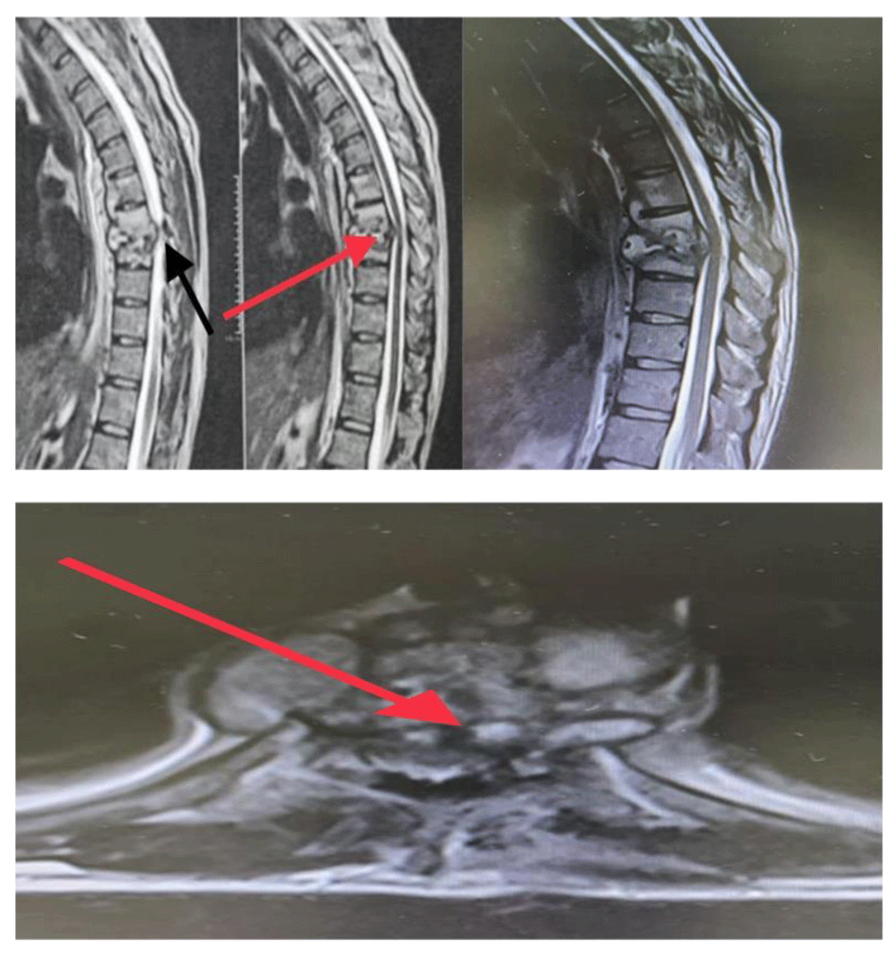
The MRI of the current case suggested a primary diagnosis of an inflammatory/infectious process probable and spondylodiscitis. The MRI (Figure 1), showed a signal alteration involving the vertebral bodies of T5, T6, T7, T8, and T9, as well as the respective disc spaces, more importantly at the levels of T6-T7 and T7-T8, presenting heterogeneous enhancement to the paramagnetic contrast agent. It is associated with bone irregularities of the corresponding apposition plates and somatic reduction in the T7 and T8 vertebral bodies' height by about 60%.
The involvement of anterior paravertebral soft tissues is characterized by densification with heterogeneous enhancement to paramagnetic contrast, delimiting areas with a low intermingled signal. It may denote the difference of the component with higher liquefaction / multiloculated collection, which extends from T5-T6 to T8-T9.
The process is expanded to the anterior and posterior epidural spaces involving the edges of the dural sac, spreading to the lateral recesses, notably in T7-T8. These two bodies have a reduction in the amplitude of the vertebral canal with compression of the adjacent spinal cord, presenting a slight change in its sign (compressive myelopathy).
Densification with heterogeneous contrast enhancement is observed involving the posterior paravertebral myoadipose planes, predominantly at the level of T7-T8 on the left. The findings described are the primary differential diagnosis of an inflammatory/infectious process (probable spondylodiscitis); however, until this moment, the laboratory data was still pending.
Incipient vertebra marginal osteophytosis (spondylosis). A good posterior alignment of vertebral bodies with conserved heights and preserved interapophyseal joints is noticed. Other intervertebral discs with preserved height and signal. There are no significant disc herniations. The rest of the vertebral canal with preserved dimensions, and the rest of the spinal cord with standard thickness and signal. Twenty-four hours later, the patient underwent a posterior percutaneous puncture of the thoracic spine (T7-T8). The IMR images suggest Spondylodiscitis in the T7-T8 levels and abscesses in the anterior and epidural regions. The bone and vertebral disc samples were sent for bacteriological identification and anatomopathological study.
Therapeutic and surgical approach
The patient remained hospitalized on a broad-spectrum antibiotic (Vancomycin + Cefepime). After the culture result with antibiogram, in which the bacterium Moraxella osloensis was identified, the infectious diseases team replaced the antibiotic with oral Rifampicin and Ciprofloxacin, and the patient was discharged from the hospital, as well as outpatient follow-up at home.
Seven days later, the patient returned, significantly worsening her general condition and pain. This situation demonstrated the inefficiency of oral antibiotic therapy for treating the pathology. A new MRI of the thoracic spine was performed. It showed an increased abscess volume in the region anterior to the T7-T8 segment discs, with extensions up to the T5 and T9 levels.
Given the worsening of the clinical picture, the patient remained hospitalized with the reintroduction of antibiotics intravenously, which is the standard gold treatment for this pathological process.
Despite this last intervention by the infectious disease team, the patient evolved clinically unsatisfactorily, showing sensory and motor deficits in the lower limbs. Therefore, urgent posterior thoracic decompression was performed (Total Laminectomy T7 and T8, with preservation of the posterior complex) to prevent severe and permanent neurological damage. It was a substantial improvement in the neurological pattern, including recovery of motor strength and the absence of paresthesia in the lower limbs.
The patient remained hospitalized, and even with intravenous antibiotic therapy directed by an antibiogram, the patient's serial imaging exams showed a progressive increase in the abscess, revealing the need for an anterior approach intervention in a mandatory and unmistakable way.
Because of the difficulty in treating the thoracic spine via the anterior route, including poor visibility and injury risk to vital structures, the thoracic surgery team was asked to access this region.
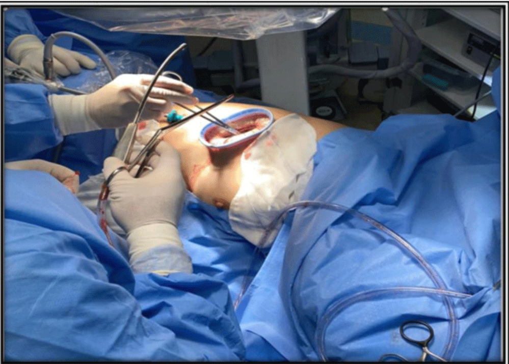
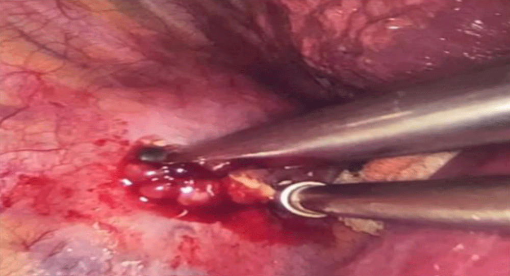
Thus, to improve the surgical outcome, a minimally invasive surgical access was conducted by video thoracoscopy (Figure 2). It is a mono-pulmonary ventilation system with a double-lumen endobronchial tube and a mini-thoracotomy with wound circumferential elastic retraction protector to maximize the treated area with minimal local trauma. After anterior decompression, the abscess was drained, and a material sample was collected for microorganism identification. Additionally, surgical debridement was performed. Then, a thoracic tubular followed the thoracic discharge and pulmonary re-expansion.
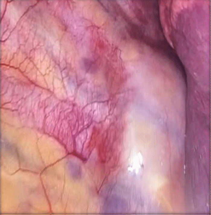
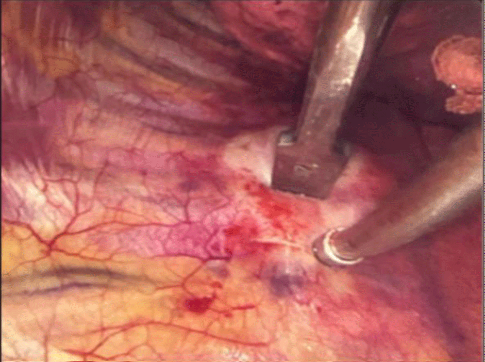
We used a pulsed lavage system with high-pressure saline solution to help debride devitalized tissues of this multiple-lobed abscess (Figures 3-5). It was intended to treat the infection and minimize deformity, preventing further surgical intervention with osteotomy and fixation by posterior approach.
In addition, we placed a local graft with bactericidal potential after debridement to assist in resolving the infectious condition and stimulate bone consolidation (Arthrodesis T7-T8).
Intraoperative Neurophysiologic Monitoring (IONM) was conducted during the surgery using the Natus neurology EDX Endeavor system. The baseline responses, with the patient at the supine position, showed TCE-MEPs and SSEPs with typical amplitudes and latencies, which were stable after positioning at left lateral decubitus. Tests were repeated after every manipulation of the vertebral body to recognize any change in neurologic status and warn the surgery team. Throughout the procedure, all responses were kept intact, indicating no injury to the medulla or other parts of the nervous system.
After performing the surgical procedure and being hospitalized, the patient waited for the culture result with Intravenous Antibiotic Therapy (Vancomycin + Cefepime).
The culture result revealed Mycobacterium tuberculosis; the treatment with Rifampicin, Isoniazid, Pyrazinamide, and Ethambutol (RIPE) was started, with therapy and outpatient follow-up scheduled for 12 months. The patient remains asymptomatic and without neurological deficit, followed up with the Infectious Diseases group in regular use of medications.
Discussion
The challenges in treating Pott's disease begin with the establishment of the correct diagnosis. It involves bacteriological or microscopic examination, culture test of the infected tissues taking into consideration findings from imaging tests, blood cultures, and histopathological analysis. MRI is also valuable for diagnosing Potts disease because it can show spinal deformities, disc destruction, or abscesses [5]. The disease can remain undiagnosed or be diagnosed later due to the lack of specific clinical manifestations involving severe neurologic complications for the patient [6], who may progress to incomplete or complete paraplegia [7].
The surgical approach was a crucial decision in managing this clinical case. The anterolateral extra pleural or the open transthoracic transpleural approach is the standard surgical method of tubercular thoracic spine decompression. It delivers satisfactory clinical outcomes but is associated with substantial morbidity and extended hospital stay [8].
In our medical experience, we observe the increasing use of minimally invasive methods to treat spinal infections, and more severe spinal lesions, with minimal damage to the soft tissue structures, rapid recovery, and early return to a functional level. Video-assisted surgery has expanded its use significantly because it has the benefits of anterior spinal surgery and provides an equivalent result of spinal deformity correction of open procedures [9].
Hence, the patient underwent a minimally invasive surgical procedure, by video laparoscopy, with anterior decompression, abscess drainage, and mechanical surgical cleaning of devitalized tissues.
During thoracic spinal manipulation, the medulla is exposed to a variety of injury mechanisms, such as ischemia due to hypotension, blood loss, stretching of the spinal cord vascular supply or segmental artery ligation; and mechanical cord compression by spinal implants, retropulsion material, hematoma and others [10]. Under these circumstances, neurophysiologic monitoring (IONM) has significantly decreased complications associated with correcting complex, severe, and rigid post-tubercular deformities [11]. Hence, we used this helpful tool [12] because it encompasses neurophysiologic tests assessing motor and sensory pathways within the spinal cord. The commonly used tests in thoracic spinal surgery are SSEP, providing information regarding dorsally located ascending spinal cord sensory tracts, and TCE-MEP, evaluating the functional properties of the anterior and lateral medulla descending corticospinal motor tracts [13].
Although drug therapy is considered the gold standard for extrapulmonary TB treatment, therapy alone is not the most appropriate in some cases. Some patients are drug-resistant, have an unsatisfactory response to treatment, or have a severe neurological compromise. Then, video-assisted surgery can help decompress the spine abscess, like in this clinical case.
The surgical approach presented in this case is based on the involvement of the vertebrae, the compressive mass, and the patient condition. The published evidence in low and middle-income countries of video-assisted thoracoscopic use is still limited, although it is a practical option for establishing thoracotomy with minimal morbidity [14].
Conclusion
Pott disease is often diagnosed during its late stages, which can be associated with devastating consequences to the patient with considerable morbidity, not to mention the associated costs. Although the same basic principles for treating pulmonary TB apply to extrapulmonary TB, drug therapy alone is sometimes inadequate; hence, the multidisciplinary approach to managing this disease can benefit the patient and improve clinical outcomes.
Declarations
Ethical approval
The patient signed a consent declaration for using images of her spine diagnostic tests to be published in journals and presentations of medical and scientific nature.
Competing interest
The authors declare no competing interests
Author’s contributions
Author Contributions: Conceptualization, AEMC; methodology, BEGD, and WHA; investigation, TEFC, and BEGD, writing—original draft preparation, all authors; writing—review and editing, all authors; visualization, LM, and RF; supervision LMRR and AEMC.
Funding
No external funding was used.
References
- WHO-a. July 6, 2022. https://www.who.int/health-topics/tuberculosis#tab=tab_1
- Moule MG, Cirillo JD. Mycobacterium tuberculosis Dissemination Plays a Critical Role in Pathogenesis. Front Cell Infect Microbiol. 2020 Feb 25;10:65. doi: 10.3389/fcimb.2020.00065. PMID: 32161724; PMCID: PMC7053427.
- World Health Organization. Global tuberculosis report 2018. WHO/CDS/TB/2018.20. Geneva: The WHO Organization; 2018.
- Viswanathan VK, Subramanian S. Pott Disease. [Updated 2022 May 1]. In: StatPearls [Internet]. Treasure Island (FL): StatPearls Publishing; 2022 Jan-Available from: https://www.ncbi.nlm.nih.gov/books/NBK538331/
- Alshoabi SA, Almas KM, Aldofri SA, Hamid AM, Alhazmi FH, Alsharif WM, Abdulaal OM, Qurashi AA, Aloufi KM, Alsultan KD, Omer AM, Daqqaq TS. The Diagnostic Deceiver: Radiological Pictorial Review of Tuberculosis. Diagnostics (Basel). 2022 Jan 25;12(2):306. doi: 10.3390/diagnostics12020306. PMID: 35204395; PMCID: PMC8870832.
- Pu F, Feng J, Yang L, Zhang L, Xia P. Misdiagnosed and mismanaged atypical spinal tuberculosis: A case series report. Exp Ther Med. 2019 Nov;18(5):3723-3728. doi: 10.3892/etm.2019.8014. Epub 2019 Sep 17. PMID: 31611930; PMCID: PMC6781804.
- Ahmed N, Khan MSI, Ahsan MK. Pott’s Paraplegia. In (Ed.), Paraplegia -New Insights [Working Title]. IntechOpen. 2022.
- Hanaoka N, Kawasaki Y, Sakai T, Nakamura T, Nanamori K, Nakamura E, Uchida K, Yamada H. Percutaneous drainage and continuous irrigation in patients with severe pyogenic spondylitis, abscess formation, and marked bone destruction. J Neurosurg Spine. 2006 May;4(5):374-9. doi: 10.3171/spi.2006.4.5.374. PMID: 16703904.
- Vaishya R, Vijay V, Agarwal AK. Video-Assisted Thoracoscopic Surgery for Drainage of Dorsal Paravertebral Abscess. Arthrosc Tech. 2017 Jul 31;6(4):e1137-e1143. doi: 10.1016/j.eats.2017.03.035. PMID: 29354409; PMCID: PMC5621866.
- Devlin VJ, Schwartz DM. Intraoperative neurophysiologic monitoring during spinal surgery. J Am Acad Orthop Surg. 2007 Sep;15(9):549-60. doi: 10.5435/00124635-200709000-00005. PMID: 17761611.
- Ruparel S, Tanaka M, Mehta R, Yamauchi T, Oda Y, Sonawane S, Chaddha R. Surgical Management of Spinal Tuberculosis-The Past, Present, and Future. Diagnostics (Basel). 2022 May 24;12(6):1307. doi: 10.3390/diagnostics12061307. PMID: 35741117; PMCID: PMC9221609.
- Eager M, Shimer A, Jahangiri FR, Shen F, Arlet V. Intraoperative neurophysiological monitoring (IONM): lessons learned from 32 case events in 2069 spine cases. Am J Electroneurodiagnostic Technol. 2011 Dec;51(4):247-63. PMID: 22303776.
- Stecker MM. A review of intraoperative monitoring for spinal surgery. Surg Neurol Int. 2012;3(Suppl 3):S174-87. doi: 10.4103/2152-7806.98579. Epub 2012 Jul 17. PMID: 22905324; PMCID: PMC3422092.
- Kapoor S, Kapoor S, Agrawal M, Aggarwal P, Jain BK Jr. Thoracoscopic decompression in Pott's spine and its long-term follow-up. Int Orthop. 2012 Feb;36(2):331-7. doi: 10.1007/s00264-011-1453-x. Epub 2012 Jan 4. PMID: 22215368; PMCID: PMC3282856.
Content Alerts
SignUp to our
Content alerts.
 This work is licensed under a Creative Commons Attribution 4.0 International License.


