Medicine Group . 2022 November 24;3(11):1393-1396. doi: 10.37871/jbres1609.
Six Examples with Different Means of Operative Treatment of Humeral Shaft Fracture
Marijo Bekic*, Marko Golubovic, Jakisa Lojpur, Nenad Sesic, Marko Moretti and Marin Pericic
- Humeral shaft fracture
- Bone healing
- Operative treatment
Abstract
We showed different cases and operative treatment of traumatic fractures of humeral shaft including operative treatment by means of external fixation, intramedullary fixation with long humeral nailing, intramedullary fixation with Rush nail and plate osteosynthesis. The indications were chosen according to fracture anatomy and biology but foremost by specific socioeconomic comorbidities and aspects of the patient itself. Bone healing status, postoperative local status, neurocirculatory status, postoperative physical therapy course and overall functional results of injured limb and patient satisfaction.
Introduction
The humerus is the largest bone in the upper extremity. Midshaft humeral fractures account for about 2-5 % of all fractures [1,2].They show bimodal distribution; the first peak is seen in the third decade in males and is associated with high-energy trauma; the second peak occurs in females in the seventh decade and is associated with low velocity falls .Trauma, increasing age, and osteoporosis are known risk factors [3-6].
This patients presents with severe arm pain in area of the mid-arm. Shortening of the upper arm suggests the presence of significant humeral shaft displacement. Evaluation of midshaft humerus fractures includes a detailed neurovascular exam of the affected arm. The radial nerve is most commonly injured by midshaft humerus fractures [1,4-6].
Fractures of the humeral shaft can be spiral, oblique or transverse. There are absolute and relative indications for surgical treatment.
Patients and Methods
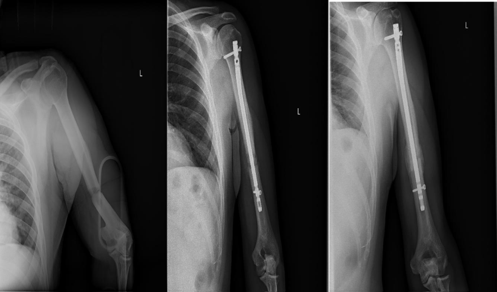
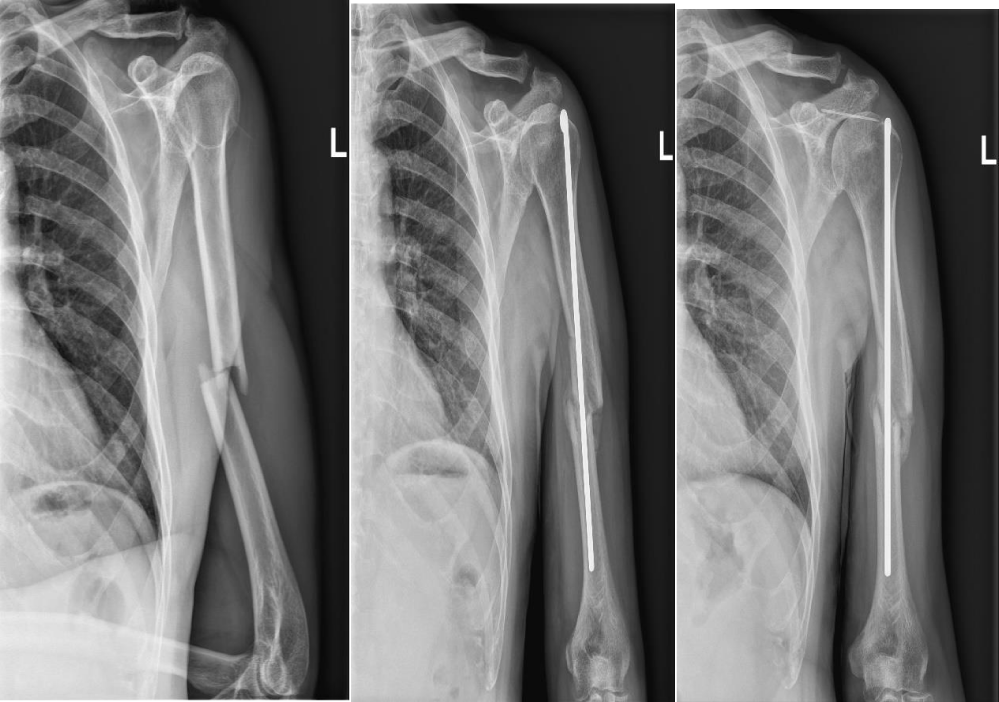
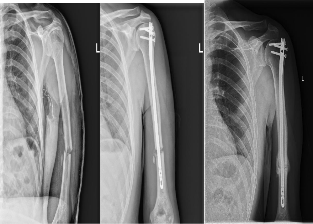
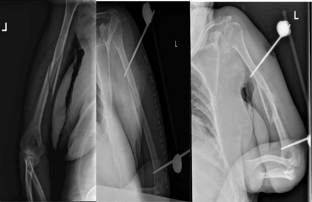
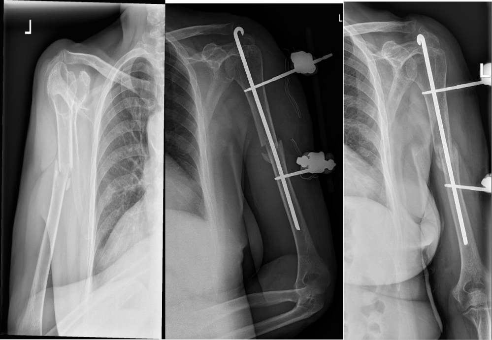
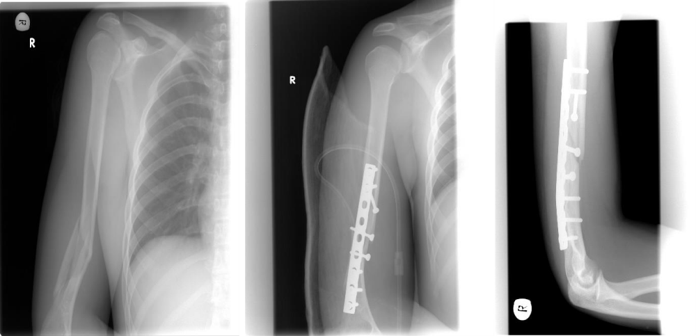
We are reporting diferent cases of operative treatment of traumatic fractures of humeral shaft including operative treatment by means of external fixation, intramedular fixation with long humeral nailing, intramedular fixation with Rush nail and plate osteosynthesis.
A study represents patients with closed midshaft humerus fractures who were treated in emergency room.
Radioraphs of the humeral shaft in an anteroposterior and lateral plane were necessary to evaluate the amount of angulation or displacement of fracture. Radiographs should include shoulder and elbow joints as well.
After evaluation of anatomy and biology of the fractures and backround of the patient and capabillities of further home care here are some cases we treated in a certain way with overall good bone healing status, postoperative local status, neurocirculatory status, postoperative physical terapy course and overall functional results of injured limb and good patient satisfaction (Figures 1-6).
Results
Follow-up is initiated on a biweekly basis until radiographic and clinical union has occured. Typical time for hard callus forming was 6 to 8 weeks.
In this study, no complications associated with humeral shaft fractures such as neurovascular compromise and nonunion were recorded. Radial nerve injury, the most common neurological complication of humeral shaft fracture that occurs in 11% of midshaft fractures didn't occur [7,8].
There was no evidence of nonunion of humeral shaft fractures. All fractures healed without infection.
Discussion
Several options are used for the management of humeral shaft fractures; conservative management, open Reduction and Internal Fixation (ORIF) with a plate, or closed reduction and Intramedullary Nailing (IMN). External fixator is also option, Most humeral shaft fractures has to be treated surgically; only small percent require functional bracing or hanging casts [9]. Today, there is an increased tendency to choose surgical management of humeral shaft fracture.The trend towards a more operative approach could be explained by the increased demand of patients and achievement of earlier immobilization.
It is generally accepted that acute, closed, uncomplicated fractures of the humeral diaphysis that occur in ambulatory, co-operative patients have high rates of union with good functional results, if treated nonoperatively. However, as for any treatment, the indications and contraindications for applying nonoperative treatment in fractures of the humeral diaphysis are constantly reviewed and subject to change, as surgical techniques are improving and the socioeconomic environment favors treatment options that can offer a faster recovery and earlier return to normal activities. Innovations in surgical techniques also play an important role.
Open reduction and internal fixation with a plate includes a 4.5 mm plate that should cover a minimum of six cortices above and below the fracture site [10]. This open operative technique allows a radial nerve identification and protection. For elderly patients, when faced with poor bone quality, there is smaller biomechanical benefit.
Intramedullary nailing can offer biomechanical and surgical advantages over plating. It allows lower bending forces and better load sharing [7,11]. Patterns in which intramedullary nailing has been found to be superior are pathological and impending fractures, segmental lesions and fractures in osteopenic bone. A simple middle transverse fracture is also a good indication for intramedularry nailing. Nail inserted through a smaller incision, allows for less soft tissue stripping compared to plating techniques.
There are many studies comparing intramedullary nailing and plating outcomes for humeral shaft fractures. One study comparing modern lacking nails with direst compression plating, fopund no significant difference regarding union rates and functional outcomes [12]. This and some other studies recorded a higher complication rate after nailing, such are shoulder impingement, restriction of shoulder movement and need for hardware removal associated with nailing [13,14]. No significant differences regarding infection, nonunion, radial nerve injury and implant failure were noticed.
External fixation is an option in cases such as polytrauma patients with severe soft tissue damage, open contaminated fracture and associated vascular injury [15-17]. Studies reporting external fixators outcomes are rare.
In our study, we showed excellent results regarding external fixator humeral shaft fracture treatment. The union rate in patients with humeral shaft fractures using external fixator was the same as in other groups.
Healing of a mid-shaft humerus fracture with surgically management shorten healing times and improve aligment compred to non-surgically treatment [5].
Invasive bridge plating through an anterior incision with functional bracing in 110 patients reported a nonunion rate of 0 % for the surgical group versus 15% for the bracing group [18].
A randomized trial involving 60 patients comparing ORIF compression plating with functional bracing reported a shorter time in union in those treated with ORIF (13.9 versus 18.7 weeks) [5,19].
Bone healing is usually divided into three slightly overlapping stages; inflammatory, reparative, and remodeling [20-23]. It is difficult to provide an approximate time frame for each phase because healing rates very widely according to age and comorbidities. Initially callus formation is cartilaginous, clinical union may occur before evidence of radiographic union is appreciable on radiographs. Clinical union classically marks the end of the reparative phase of fracture healing.
Some conditions impair fracture healing rates such as patient age, comorbidities, diabetes mellitus, arteriovascular disease, anemia, hypothyrodism, malnutrition, excessive chronic alcohol and tobacco use. Some medications impair fracture healing [24]. Our study showed no difference among patients. Health status and age showed no difference in bone healing.
Plain radioraphs are sufficient to confirm the diagnosis and plan the treatment for humeral shaft fractures. Anteroposterior and lateral radiographs are required to visualize and make a full assessment of the fracture.
In our cases, there was no difference concering functional outcomes between different operative and surgical management for humeral shaft fractures. Therefore, an humeral shaft fracture is proper indication for operative and surgical management.
Most cases of fixation failure can be attributed to poor technique such as too a short plate, but biomechanical studies showed that a plate restore excellent bending and torsional stiffness to the humerus [25-28].
Competent fracture care requires a basic knowledge of bone biology and healing. Practical understanding and managing fractures is necessary.
References
- Carroll EA, Schweppe M, Langfitt M, Miller AN, Halvorson JJ. Management of humeral shaft fractures. J Am Acad Orthop Surg. 2012 Jul;20(7):423-33. doi: 10.5435/JAAOS-20-07-423. PMID: 22751161.
- Court-Brown CM, Caesar B. Epidemiology of adult fractures: A review. Injury. 2006 Aug;37(8):691-7. doi: 10.1016/j.injury.2006.04.130. Epub 2006 Jun 30. PMID: 16814787.
- Ekholm R, Adami J, Tidermark J, Hansson K, Törnkvist H, Ponzer S. Fractures of the shaft of the humerus. An epidemiological study of 401 fractures. J Bone Joint Surg Br. 2006 Nov;88(11):1469-73. doi: 10.1302/0301-620X.88B11.17634. PMID: 17075092.
- Lineage Medical, Inc., OrthoBullets. Humeral Shaft Fractures. National Center for Biotechnology Information, StatPearls. Humeral Shaft Fractures. 2021.
- Tytherleigh-Strong G, Walls N, McQueen MM. The epidemiology of humeral shaft fractures. J Bone Joint Surg Br. 1998 Mar;80(2):249-53. doi: 10.1302/0301-620x.80b2.8113. PMID: 9546454.
- Ekholm R, Ponzer S, Törnkvist H, Adami J, Tidermark J. Primary radial nerve palsy in patients with acute humeral shaft fractures. J Orthop Trauma. 2008 Jul;22(6):408-14. doi: 10.1097/BOT.0b013e318177eb06. PMID: 18594306.
- Ekholm R, Tidermark J, Törnkvist H, Adami J, Ponzer S. Outcome after closed functional treatment of humeral shaft fractures. J Orthop Trauma. 2006 Oct;20(9):591-6. doi: 10.1097/01.bot.0000246466.01287.04. PMID: 17088659.
- Clement ND. Management of Humeral Shaft Fractures; Non-Operative Versus Operative. Arch Trauma Res. 2015 Jun 20;4(2):e28013. doi: 10.5812/atr.28013v2. PMID: 26401493; PMCID: PMC4577941.
- Updegrove GF, Mourad W, Abboud JA. Humeral shaft fractures. J Shoulder Elbow Surg. 2018 Apr;27(4):e87-e97. doi: 10.1016/j.jse.2017.10.028. Epub 2017 Dec 29. PMID: 29292035.
- Hughes RE, Schneeberger AG, An KN, Morrey BF, O'Driscoll SW. Reduction of triceps muscle force after shortening of the distal humerus: a computational model. J Shoulder Elbow Surg. 1997 Sep-Oct;6(5):444-8. doi: 10.1016/s1058-2746(97)70051-x. PMID: 9356933.
- Putti AB, Uppin RB, Putti BB. Locked intramedullary nailing versus dynamic compression plating for humeral shaft fractures. J Orthop Surg (Hong Kong). 2009 Aug;17(2):139-41. doi: 10.1177/230949900901700202. PMID: 19721138.
- Kurup H, Hossain M, Andrew JG. Dynamic compression plating versus locked intramedullary nailing for humeral shaft fractures in adults. Cochrane Database Syst Rev. 2011 Jun 15;(6):CD005959. doi: 10.1002/14651858.CD005959.pub2. PMID: 21678350.
- Ouyang H, Xiong J, Xiang P, Cui Z, Chen L, Yu B. Plate versus intramedullary nail fixation in the treatment of humeral shaft fractures: an updated meta-analysis. J Shoulder Elbow Surg. 2013 Mar;22(3):387-95. doi: 10.1016/j.jse.2012.06.007. Epub 2012 Sep 1. PMID: 22947239.
- Marsh JL, Mahoney CR, Steinbronn D. External fixation of open humerus fractures. Iowa Orthop J. 1999;19:35-42. PMID: 10847515; PMCID: PMC1888611.
- Mostafavi HR, Tornetta P 3rd. Open fractures of the humerus treated with external fixation. Clin Orthop Relat Res. 1997 Apr;(337):187-97. doi: 10.1097/00003086-199704000-00021. PMID: 9137190.
- Zinman C, Norman D, Hamoud K, Reis ND. External fixation for severe open fractures of the humerus caused by missiles. J Orthop Trauma. 1997 Oct;11(7):536-9. doi: 10.1097/00005131-199710000-00013. PMID: 9334957.
- Matsunaga FT, Tamaoki MJ, Matsumoto MH, Netto NA, Faloppa F, Belloti JC. Minimally Invasive Osteosynthesis with a Bridge Plate Versus a Functional Brace for Humeral Shaft Fractures: A Randomized Controlled Trial. J Bone Joint Surg Am. 2017 Apr 5;99(7):583-592. doi: 10.2106/JBJS.16.00628. PMID: 28375891.
- Hosseini Khameneh SM, Abbasian M, Abrishamkarzadeh H, Bagheri S, Abdollahimajd F, Safdari F, Rahimi-Dehgolan S. Humeral shaft fracture: a randomized controlled trial of nonoperative versus operative management (plate fixation). Orthop Res Rev. 2019 Sep 23;11:141-147. doi: 10.2147/ORR.S212998. PMID: 31576178; PMCID: PMC6765056.
- Wilkins KE. Principles of fracture remodeling in children. Injury. 2005 Feb;36 Suppl 1:A3-11. doi: 10.1016/j.injury.2004.12.007. PMID: 15652934.
- Jones ET. Skeletal growth and development as related to trauma. In: Skeletal Trauma in Children, 3rd, Green NE, Swiontkowski MF (Eds), Saunders, Philadelphia 2003. p.6.
- Dimitriou R, Tsiridis E, Giannoudis PV. Current concepts of molecular aspects of bone healing. Injury. 2005 Dec;36(12):1392-404. doi: 10.1016/j.injury.2005.07.019. Epub 2005 Aug 15. PMID: 16102764.
- Buckwalter JA, Glimcher MJ, Cooper RR, Recker R. Bone biology. I: Structure, blood supply, cells, matrix, and mineralization. Instr Course Lect. 1996;45:371-86. PMID: 8727757.
- Gaston MS, Simpson AH. Inhibition of fracture healing. J Bone Joint Surg Br. 2007 Dec;89(12):1553-60. doi: 10.1302/0301-620X.89B12.19671. PMID: 18057352.
- Zimmerman MC, Waite AM, Deehan M, Tovey J, Oppenheim W. A biomechanical analysis of four humeral fracture fixation systems. J Orthop Trauma. 1994;8(3):233-9. doi: 10.1097/00005131-199406000-00009. PMID: 8027893.
- Lin J, Inoue N, Valdevit A, Hang YS, Hou SM, Chao EY. Biomechanical comparison of antegrade and retrograde nailing of humeral shaft fracture. Clin Orthop Relat Res. 1998 Jun;(351):203-13. PMID: 9646764.
- Blum J, Machemer H, Baumgart F, Schlegel U, Wahl D, Rommens PM. Biomechanical comparison of bending and torsional properties in retrograde intramedullary nailing of humeral shaft fractures. J Orthop Trauma. 1999 Jun-Jul;13(5):344-50. doi: 10.1097/00005131-199906000-00004. PMID: 10406701.
- Henley MB, Monroe M, Tencer AF. Biomechanical comparison of methods of fixation of a midshaft osteotomy of the humerus. J Orthop Trauma. 1991;5(1):14-20. doi: 10.1097/00005131-199103000-00003. PMID: 2023038.
- Rockwood and Green’s: Fractures in Adults; Humeral shaft fractures. 2015.
Content Alerts
SignUp to our
Content alerts.
 This work is licensed under a Creative Commons Attribution 4.0 International License.


