Medicine Group. 2024 January 17;5(1):044-051. doi: 10.37871/jbres1870.
Risk Factors for Early Bioprosthetic Heart Valve Degeneration in Patients with Rheumatic Fever
Giovanna Paliares Monteiro1, Carolina Carotenuto Ramos1, Livia Airoldi Broekman1, Aldrei Costa Araujo1 and Jean Marcos de Souza1,2*
2Division of Rheumatology, Universidade Estadual de Campinas and Universidade de Sao Paulo, Brazil
- Rheumatic fever
- Rheumatic heart disease
- Bioprosthesis
- Valve replacement
Abstract
Background: Rheumatic Fever (RF) is an immunological disorder related to exposure to group a streptococcus and rheumatic heart disease is an important cause of valve replacement. Bioprosthetic valves tend to degenerate faster in rheumatic patients, presumably due to immune mechanisms.
Objectives: The study sought to assess whether classic risk factors for cardiovascular disease are related to Early Valve Degeneration (EVD) in patients with RF.
Design: Case-control study.
Methods: Patients with RF and EVD or Late Valve Degeneration (LVD) were selected. The cutoff point was 9 years for a second valve surgery. Data regarding cardiovascular risk of the two groups were obtained and compared. A data imputation analysis was used to deal with missing data.
Results: Twenty valves were included in the primary outcome analysis and 33 were used for data imputation. The mean age for the first valve replacement was 40.6 (±6.2) years for the EVD group and 31.1 (±12.3) years for the LVD group, (p = 0.03), which remained significant after data imputation. For blood pressure, there was a non-statistically significant trend towards higher diastolic pressure in patients with EVD in relation to LVD (86.8 (±7.2) and 79 (±10.9) mmHg, respectively, p = 0.08), which after data imputation was statistically higher than that of the LVD group (88 [85.4-88.8] and 77 [73-84.5] mmHg, respectively, p = 0.001). Lipid profile was also worse on the EVD group.
Conclusions: The data suggest that EVD may result more from aging and cardiovascular factors than from immunological mechanisms, suggesting stricter targets for cardiovascular disease in these patients.
Introduction
Rheumatic Fever (RF) is an immune disorder related to repetitive Group A Streptococcal (GAS) exposure. Among its chronic manifestations, rheumatic heart disease (RHD) is the most relevant, because it is an important cause of structural heart disease [1]. Mitral valve disease is the most common (and mitral valve stenosis is very characteristic of RF), followed by the aortic valve [2]. The chronic mitral valve insufficiency of rheumatic ethology, occasioned by structural modifications that results in failure of the valve, predominates in the young and is an important factor for valve replacement [3]. In the elderly, the valve calcification associated to aortic stenosis is the main indication for valve surgery. Tricuspid insufficiency and stenosis can also require valve replacement [4]. Because of the relatively young age of valve replacement surgery, it is important to investigate intrinsic risk factors when choosing a valve (biological, mechanical), as well as the organism of the receiving patient that leads to contention or aggravation of the prosthesis degeneration.
There is limited evidence for pharmacological treatment for RHD and the valve replacement is still the only definite treatment [5,6]. Nowadays, two major groups of prothesis are available at the market: the mechanical valve and the biological (bioprosthesis) valve [7]. The choice of which valve will be used is individualized for each patient, considering mainly age and associated comorbidities. Guidelines recommend bioprosthesis for older patients, usually over 70 years old, while the mechanical valves are recommended to younger patients, usually below 50 years old. For patients between 50-70 years old, there is no preference [8]. It is known that the mechanical prosthesis last longer than the bioprosthesis, as there is a lower chance of Structural Valve Degeneration (SVD), which reduces the necessity of reoperation. Nonetheless, they lead to an increased risk of endocarditis, thromboembolic, and cerebrovascular events when compared to the bioprosthesis, requiring permanent anti-coagulation, increasing the changes of hemorrhages and cerebrovascular events [6]. Studies by Hamamoto and colleagues compare the durability of biological prosthesis in rheumatic patients and non-rheumatic patients and the necessity of long-term reoperation [9]. They have shown that the bioprosthetic valve survival in patients with no RHD were 85%, 76% and 63% in 5, 10 and 15 years, respectively; meanwhile, for patients with RHD, the results were 89%, 46% and 5% in 5, 10 and 15 years [9].
That being said, it is plausible that the time frame for valve replacement can be individualized and depends on what kind of prothesis will be chosen, the age of the patient, their comorbidities and life style. Diseases such as hypertension, diabetes mellitus, atherosclerosis, dyslipidemia, renal insufficiency and cardiac failure are the main conditions that, allegedly, when controlled, can lead to a better overall survival and might decrease the need of a valve replacement, as such risk factors can precipitate multiple forms of early valve damage, as calcifications, fibrosis, thrombosis or endocarditis [10].
Thus, our study aims to analyze if there is correlation between classical cardiovascular risk factors and early bioprosthetic degeneration in patients with RF, considering most conditions previously mentioned are, in general, avoidable with interventions other than immune modulation.
Materials and Methods
Study design
This case-control study selected RF patients with Early Valve Degeneration (EVD) Or Late Valve Degeneration (LVD), according to the criteria further described. Through medical reports and interviews, data regarding the cardiovascular risk during the first valve replacement, during the inter-operative period and during the re-operation period or re-operation indication (for those awaiting surgery still), was obtained. Data were submitted for analysis in order to establish differences regarding the risk factors associated with the degeneration pattern.
Selection
The selected patients were followed at the outpatient clinic of the Rheumatology division from Hospital das Clinicas of Universidade de Sao Paulo, Brazil. Inclusion criteria were: patients with RF diagnosed according to Jones criteria revised in 2015 by the American Heart Association [11], both genders, older than 18 years old, previous valve replacement for a biological prosthesis due to RHD, and: 1) at least one re-operation due to biological prosthesis degeneration; or 2) awaiting in line for heart surgery due to biological prosthesis degeneration. Patients who underwent more than one replacement of the bioprosthetic valve were assessed multiple times.
Exclusion criteria were: other autoimmune diseases, neoplasia at any moment, surgical failure during first surgery or during the surgery intended for this analysis culminating in valve replacement not due to degeneration but on account of prothesis dysfunction associated to surgical failure, infectious endocarditis, or new rheumatic episode well described during analysis.
Recruited patients were divided in 2 groups: EVD and LVD, according to the criteria of 9 years until necessity of new surgery, defined by the authors regarding Hamamoto's study [9]. It was shown in the study that the time frame for new surgical intervention for 50% of the analyzed rheumatic fever patients is, approximately, of 9 years.
This study was approved by the Ethics Committee and the informed writing consent was signed by all patients. The study was conducted in accordance to Brazilian resolution 466/2012.
Clinical and laboratory evaluation
In a longitudinal observation, all patients had their electronic medical reports accessed to gather the following data: age, sex, blood pressure, High-Density Lipoprotein (HDL), Low-Density Lipoprotein (LDL), triglycerides, glucose, creatinine and glycosylated hemoglobin (HbA1c).
By using electronic questionnaire, answered remotely and in person during outpatient follow ups, data such as penicillin use and compliance, ethnicity, and atherosclerosis history was collected.
Data was collected regarding the variables “age”, “sex”, “ethnicity” and “penicillin use” with reference to the first surgery — or the surgery concerning the analysis, in case the patient in question was submitted to multiple surgeries.
As for the variables “penicillin use compliance”, the approximate number of lost applications per year was obtained according to patient’s report; for “blood pressure”, the mean Systolic Blood Pressure (SBP) and Diastolic Blood Pressure (DBP) were obtained according to medical report’s values during the analyzed period; for laboratory serial values, it was used means regarding the period analyzed.
With reference to “atherosclerosis history”, the dichotomous answer was considered regarding acute myocardial infarction, ischemic atherothrombotic stroke or atherosclerotic peripheral vascular disease throughout the whole life until the data of analysis. In case of doubt about the reported event being caused by embolic thrombosis or atrial fibrillation, the event was not considered positive.
Statistical Analysis
Data was presented as mean ± standard deviation or median and interquartile range. The Shapiro-Wilk test was applied, verifying the sample’s normality. The categorical variables were analyzed by the Fisher test or chi-square test. The normally distributed variables were evaluated by the Student’s t-test and the not normally distributed by the Mann-Whitney’s test. The statistical power of the data to detect differences between groups was also calculated for the main outcomes [12]. Missing completely at random data were input by Expectation-Maximization (EM) algorithm. p values < .05 were considered statistically significant. For the analysis, SPSS software — version 25 (IBM, Chicago, IL, USA) was utilized.
Results
Selection of patients
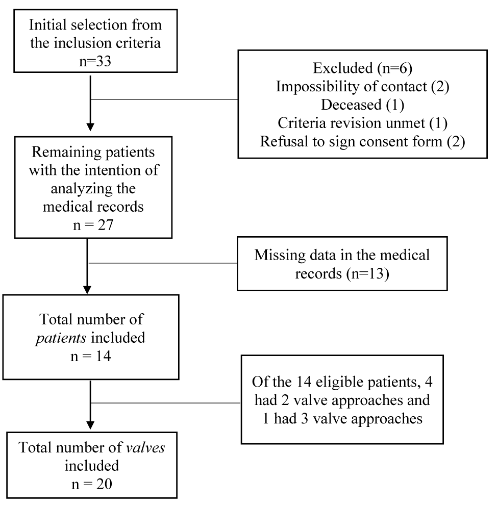
The protocol was applied from September 2021 to February 2022. The application of the inclusion criteria allowed the initial selection of 33 patients. After excluding 6 patients (two due to impossibility of contact, one due to death from SARS-CoV-2, one due to not meeting the criteria for rheumatic fever and 2 due to refusal to sign the consent form), 27 potential candidates were listed. As 4 patients had 2 valve approaches and 1 patient had 3 approaches, therefore, 33 valves were included for medical record analysis.
In 13 objects of study, the scarcity of data from medical records made it impossible to carry out the complete analysis proposed, and only 20 valves were effectively included in the study. An analysis of the 33 cases will be described below, in the section “Missing Data”, in which imputation strategies were implemented to estimate possible correlations. A schematic of the inclusion process can be found in figure 1.
Patient characteristics
Of the 20 objects of study, 6 were classified as EVD and 14 as LVD. The mean age at baseline valve replacement was 40.6 (±6.2) years for the EVD group, with the proportion of men and women being 1:5, respectively. In the LVD group, the mean age was 31.1 (±12.3) years with a ratio of men and women of 2:5. Age was significantly different between groups (p = 0.03), with a difference of almost 10 years (Table 1). The power calculation for the outcome yielded a value of 62.9%, suggesting an underpowered sample.
| Table 1: Comparison between demographic, clinical and laboratory data of patients with early and late valve degeneration. | |||
| EVD (n = 6) | LVD (n = 14) | p (95% IC) | |
| Male, % | 17 | 29 | 0.63 |
| Caucasian ethnicity, % | 50 | 57 | 1 |
| Age, years* | 40.6 (6.2) | 31.1 (12.3) | 0.03† |
| Prophylactic antibiotic, %* | 50 | 70 | 0.72 |
| Adherence to prophylaxis ‡ | 0 [0 - 4] | 3 [0 - 4] | N/A§ |
| SBP, mmHg | 127.3 (17.9) | 125.1 (14.1) | 0.91 |
| DBP, mmHg | 86.8 (7.2) | 79 (10.9) | 0.08 |
| Manifested atherosclerosis, % | 34 | 21 | 0.66 |
| HDL, mg/dL | 43.8 [42.1 - 66.7] | 50.9 [42.9 - 59.9] | 0.84 |
| LDL, mg/dL | 115 [92.1 - 126.6] | 124.5 [101 - 129.9] | 0.77 |
| TG, mg/dL | 98.1 [87.5 - 130.7] | 100.6 [90.8 - 140.5] | 0.9 |
| VLDL, mg/dL | 19.6 [18.3 - 25.6] | 20 [17.6 - 22.8] | 1 |
| Blood glucose, mg/dL | 121.1 [114.5 - 133.9] | 103 [89 - 124.2] | 0.14 |
| HbA1c, % | 5.6 [5.2 - 5.7] | 5.3 [5.1 - 5.6] | 0.62 |
| Creatinine, mg/dL | 0.9 [0.8 - 1.14] | 0.9 [0.7 - 1.1] | 0.88 |
| Data presented as mean (Standard Deviation) or median [Interquartile Range], unless otherwise specified. Abbreviations: EVD: early valve degeneration; LVD: late valve degeneration; CI: confidence interval; SBP: Systolic Blood Pressure; DBP: Diastolic Blood Pressure; HDL: High Density Lipoprotein; LDL: Low Density Lipoprotein; TG: Triglycerides; VLDL: Very Low-Density Lipoprotein; Hba1c: Glycated Hemoglobin * at the time of valve replacement used as baseline † statistically significant (with 95% confidence interval) ‡ in number of missed doses per year § statistical calculation not applicable (only 3 patients in the early arm) |
|||
Caucasian ethnicity predominated in the LVD group, with 8 patients (57%) declaring themselves white and the other 6 (43%) declaring themselves belonging to other ethnicities (African-American, Indigenous or Asiatic). In the EVD group, 3 (50%) declared themselves to be Caucasian and the other 3 (50%) of other ethnicities. However, there was no statistical difference between the groups (Table 1).
Regarding the use of penicillin during the time of heart valve replacement surgery, 3 (50%) of the patients in the EVD group used the medication and 3 (50%) did not undergo treatment with the medication in question. Among those using penicillin, the median number of missed doses per year was zero. In the LVD group, 10 volunteers (70%) used the drug and 4 of them (30%) not. Non-adherence to penicillin corresponded to a median of 3 doses per year. Neither prescription nor adherence to penicillin were statistically different between groups (Table 1).
Cardiovascular risk
Regarding the variables related to cardiovascular risk, a mean SBP and DBP of 127.3 (± 17.9) mmHg and 86.8 (± 7.2) mmHg were obtained, respectively, for the DVP group, while for the LVD group, the mean was 125.1 (± 14.1) mmHg and 79 (± 10.9) mmHg (Table 1). There was a non-significant trend (p = 0.08) for an increase in DBP in patients with EVD that was significantly accentuated in the analysis with data imputation (further described).
The occurrence of atherosclerotic events was 34% for the EVD group, with 2 patients presenting at least one of the aforementioned events. For the LVD group, 4 patients (21%) had a history of manifested atherosclerosis at some point, with no statistical difference (Table 1).
For the variable serum lipid profile, the median obtained for the EVD group was 43.8 [42.1-66.7], 115 [92.1-126.6], 98.1 [87.5-130.7], 19.6 [18.3-25.6] for HDL, LDL, triglycerides and VLDL, respectively. As for the LVD, the values were 50.9 [42.9-59.9], 124.5 [101-129.9], 100.6 [90.8-140.5], 20 [17, 6-22.8], respectively. No parameter was significantly different between groups. The median creatinine for both groups was 0.9.
Finally, the glycemic profile was evaluated through fasting glucose and HbA1c. For the EVD group, the median values were 121.1 [114.5-133.9] for blood glucose and 5.6 [5.2-5.7] for HbA1c. For the LVD group, the median blood glucose and HbA1c were 103 [89-124.2] and 5.3 [5.1-5.6], respectively. Despite the differences between blood glucose values, they were not statistically significant. The overall statistical power of the data was low, approximately 40%, indicating an underpowered sample.
Missing data
Missing data referred to blood pressure measurements and laboratory data, which could not be obtained from very old medical records. Approximately 36% of blood pressure values and 25% of laboratory test values could not be obtained. As Little's chi-square test for randomness demonstrated that the sample had completely random missing data (p = 0.9), we proceeded with EM technique, which allowed us to compile the data shown in table 2.
| Table 2: Exploratory and hypothetical comparison between groups after data imputation. | |||
| EVD (n = 14) | LVD (n = 19) | p (95% IC) | |
| Male, % | 14 | 21 | 0.68 |
| Caucasian ethnicity, % | 43 | 47 | 1 |
| Age, years* | 37 [33.2 - 40.7] | 26 [22 - 30] | 0.02† |
| Prophylactic antibiotic, %* | 64 | 78 | 0.44 |
| Adherence to prophylaxis‡ | 0 [0 - 2] | 2 [0 - 4] | 0.34 |
| SBP, mmHg | 121 [115.2 - 126.3] | 121 [114.4 - 129] | 0.7 |
| DBP, mmHg | 88 [85.4 - 88.8] | 77 [73 - 84.5] | 0.001† |
| Manifest atherosclerosis, % | 21 | 15 | 1 |
| HDL, mg/dL | 40.3 [35.3 - 44.7] | 54 [44 - 60.8] | 0.02† |
| LDL, mg/dL | 118 (15.3) | 116 (20.5) | 0.77 |
| TG, mg/dL | 216 (61.3) | 111 (41.6) | < 0.01† |
| Blood glucose, mg/dL | 100 (3.1) | 110 (18.6) | 0.14 |
| HbA1c, % | 5.6 [5.2 - 5.7] | 5.3 [5.1 - 5.6] | 0.05 |
| Data presented as mean (Standard Deviation) or median [Interquartile Range], unless otherwise specified. Abbreviations: EVD: Early Valve Degeneration; LVD: Late Valve Degeneration; CI: Confidence Interval; SBP: Systolic Blood Pressure; DBP: Diastolic Blood Pressure; HDL: High Density Lipoprotein; LDL: Low Density Lipoprotein; TG: Triglycerides; VLDL: Very Low-Density Lipoprotein; Hba1c: Glycated Hemoglobin * at the time of valve replacement used as baseline † statistically significant (with 95% confidence interval) ‡ in number of missed doses per year |
|||
Patient characteristics (exploratory)
As shown in table 2, after compiling the random missing data, 33 study objects were selected, of which 14 were included in the EVD group (n = 14) and 19 in the LVD group (n = 19). In the EVD group, 2 patients were male, while in the LVD group, 4 were male. Being male was not statistically significant between groups (p = 0.68).
Regarding ethnicity, 6 patients declared themselves to be Caucasian in the EVD group (n = 6), whereas in the LVD group, 9 were Caucasian (n = 9). As well as being male, ethnicity also did not present significant differences between the groups. For the age variable, in years, the median was 37 [33.2-40.7] for the EVD group and 26 [22-30] for the LVD, showing a statistically significant difference between the groups (p = 0.02).
Regarding the use of prophylactic antibiotics, in the EVD group, only 9 received prophylactic antibiotic therapy (n = 9). In the LVD group, 15 patients received the medication. The use and compliance of prophylactic antibiotics were not statistically significant in the sample.
Cardiovascular risk (exploratory)
Regarding the variables related to cardiovascular risk, a median SBP and DBP of 121 [115.2-126.3] mmHg and 88 [85.4-88.8] mmHg, respectively, were obtained for the EVD group, while for the LVD group, the median was 121 [114.4-129] mmHg and 77 [73-84.5] (Table 2). Only DBP were statistically different (p = 0.001).
The occurrence of atherosclerotic events was 21% for the EVD group, while in the LVD group 15% of the patients had a history of manifested atherosclerosis at some point (Table 2), with no difference between the groups.
For the variable serum lipid profile, the mean or median obtained for the EVD group was 40.3 [35.3-44.7], 118 (± 15.3), 216 (± 61.3) for HDL, LDL and triglycerides, respectively. As for the LVD, the values were 54 [44-60.8], 116 (± 20.5), 111 (± 41.6), respectively. Only the values of HDL (p = 0.02) and triglycerides (P < 0.01) were significantly different between groups.
The glycemic profile, evaluated through fasting glucose and HbA1c, in the EVD group, had a mean of 100 (± 3.1) for glycemia and a median of 5.6 [5.2-5.7] for HbA1c. For the LVD group, the values for blood glucose and HbA1c were 110 (± 18.6) and 5.3 [5.1-5.6], respectively. HbA1c values showed a non-significant trend towards difference (p = 0.05).
After imputation of data, the statistical power of the main outcomes increased to approximately 70%, although the sample remained underpowered.
Discussion
In this case-control, older age at first valve replacement was the main factor associated with shorter bioprosthetic duration. The other cardiovascular risk factors were not significantly different between the groups.
Acquired valvular heart disease has some important risk factors such as age, sex, smoking, hypercholesterolemia, rheumatic heart disease and hypertension [13]. Among them, the mechanism of the pathogenesis of rheumatic valvular heart disease stands out, which occurs primarily by an aggression to the endocardial surface mediated by antibodies produced in response to GAS infection. They cause endothelial activation with upregulated expression of Vascular Cell Adhesion Protein 1 (VCAM1), which facilitates the adhesion of T cells to the surface of heart valves. Microscopically, subendothelial collagen and perivascular tissue are the sites most affected by the intense inflammatory process that, mediated by pro-inflammatory substances such as tumor necrosis factor, interleukin 1-β, reactive oxygen species, advanced glycosylation end products and oxidized Low-Density Lipoprotein (oxLDL) cholesterol activate biomineralization and vascular osteogenic signaling processes, which ultimately cause valvular fibrosis [13,14].
It is observed that the valve degeneration process includes a primary inflammatory component, particularly noticeable in individuals with RF. Additionally, there are non-immune processes within the patient, largely influenced by oxidative stress [13,14]. Consequently, when considering the causes of non-rheumatic valve disease, advanced age emerges as a shared risk factor across all cases [15]. The findings of this study may suggest that patients with RF experience not only direct inflammatory effects on the valve but also a potentially accelerated aging-related process due to a higher inflammatory threshold.
One of the main limitations of our study is the small number of patients included in the final analysis, which significantly influenced the statistical power of the data, potentially obscuring differences between the groups. Despite the sample’s low power, age differences were still statistically significant. In an exploratory way, we tried to impute missing data and obtained differences in the values of DBP and lipids (HDL and triglycerides). The data suggest that lower diastolic blood pressure values could be related to an increase in prosthesis preservation time and, thus, a lower need for surgical re-approach of the patient. Furthermore, laboratory values related to the cardiovascular profile (lipid and glycemic) could also impact on prosthetic survival, confirming that a better laboratory control of these parameters would positively influence the need for valve re-approach. Still, due to the large amount of missing data, all this analysis is only speculative.
A second important limitation of the study refers to the retrospective nature of the survey, subject to patient records and data from medical records. In this sense, adherence to treatment, apparently very good among the participants, may not be effectively true, causing distortions in the interpretation of results. Prospective cohort studies with prophylaxis application verification protocols (through application booklet or digital verification system) would likely help to reduce these biases.
Additionally, the small number of patients did not allow a multivariate analysis, which is so important in observational studies. It is possible that confounding factors are explaining the observed phenomenon, such as, for example, the surgical technique used or even the moment when the surgery was performed (with older patients using older techniques and using less of the service's experience). Finally, examining the immune profile of the patients, including serum interleukins, might have helped confirm our findings. This avenue could be explored in future research endeavors.
In summary, we understand that the present study has several limitations but, as far as our knowledge allows us to say, we believe that it is the first to try to identify risks of bioprosthetic degeneration in patients with RF. New prospective studies are needed and welcome, in a partially neglected clinical entity.
Conclusion
The data found in this study suggest that RF-related EVD may be more related to the age at which the valve was approached and to hemodynamic factors than to immune factors, suggesting that stricter targets for blood pressure control and cardiovascular risk factors are imperative in these patients.
Declarations
Ethics approval and consent to participate/for publication
The study was approved by the institutional Ethics Committee and all patients signed a written consent form before entering the study.
Author Contributions
The authors contributed to the manuscript as further described:
Conceptualization, J.M.S.; Methodology, J.M.S.; Investigation, G.P.M., C.C.R., L.A.B., A.C.A., and J.M.S.; Writing – Original Draft, G.P.M., C.C.R., L.A.B., A.C.A., and J.M.S.; Writing – Review & Editing, G.P.M., C.C.R., L.A.B., A.C.A., and J.M.S.; Supervision, J.M.S.
Acknowledgements
Not applicable.
Funding
We thank Conselho Nacional de Desenvolvimento Cientifico e Tecnologico (CNPq) for the scholarships granted to A.C.A.
Competing Interests
All authors declare no conflict of interest.
Availability of Data
All data will be available in reasonable time upon request to the authors.
References
- Russell EA, Tran L, Baker RA, Bennetts JS, Brown A, Reid CM, Tam R, Walsh WF, Maguire GP. A review of valve surgery for rheumatic heart disease in Australia. BMC Cardiovasc Disord. 2014 Oct 2;14:134. doi: 10.1186/1471-2261-14-134. PMID: 25274483; PMCID: PMC4196004.
- John S, Bashi VV, Jairaj PS, Muralidharan S, Ravikumar E, Sathyamoorthy I, Babuthaman C, Krishnaswamy S, Cherian G, Sukumar IP. Mitral valve replacement in the young patient with rheumatic heart disease. Early and late results in 118 subjects. J Thorac Cardiovasc Surg. 1983 Aug;86(2):209-16. PMID: 6876857.
- Gabbay U, Yosefy C. The underlying causes of chordae tendinae rupture: a systematic review. Int J Cardiol. 2010 Aug 20;143(2):113-8. doi: 10.1016/j.ijcard.2010.02.011. Epub 2010 Mar 7. PMID: 20207434.
- Tarasoutchi F, Montera MW, Ramos AIO, Sampaio RO, Rosa VEE, Accorsi TAD, Santis A, Fernandes JRC, Pires LJT, Spina GS, Vieira MLC, Lavitola PL, Ávila WS, Paixão MR, Bignoto T, Togna DJD, Mesquita ET, Esteves WAM, Atik F, Colafranceschi AS, Moises VA, Kiyose AT, Pomerantzeff PMA, Lemos PA, Brito Junior FS, Weksler C, Brandão CMA, Poffo R, Simões R, Rassi S, Leães PE, Mourilhe-Rocha R, Pena JLB, Jatene FB, Barbosa MM, Abizaid A, Ribeiro HB, Bacal F, Rochitte CE, Fonseca JHAPD, Ghorayeb SKN, Lopes MACQ, Spina SV, Pignatelli RH, Saraiva JFK. Update of the Brazilian Guidelines for Valvular Heart Disease - 2020. Arq Bras Cardiol. 2020 Oct;115(4):720-775. English, Portuguese. doi: 10.36660/abc.20201047. PMID: 33111877; PMCID: PMC8386977.
- Ralph AP, Noonan S, Wade V, Currie BJ. The 2020 Australian guideline for prevention, diagnosis and management of acute rheumatic fever and rheumatic heart disease. Med J Aust. 2021 Mar;214(5):220-227. doi: 10.5694/mja2.50851. Epub 2020 Nov 15. PMID: 33190309.
- Kumar RK, Antunes MJ, Beaton A, Mirabel M, Nkomo VT, Okello E, Regmi PR, Reményi B, Sliwa-Hähnle K, Zühlke LJ, Sable C; American Heart Association Council on Lifelong Congenital Heart Disease and Heart Health in the Young; Council on Cardiovascular and Stroke Nursing; and Council on Clinical Cardiology. Contemporary Diagnosis and Management of Rheumatic Heart Disease: Implications for Closing the Gap: A Scientific Statement From the American Heart Association. Circulation. 2020 Nov 17;142(20):e337-e357. doi: 10.1161/CIR.0000000000000921. Epub 2020 Oct 19. Erratum in: Circulation. 2021 Jun 8;143(23):e1025-e1026. PMID: 33073615.
- Harky A, Suen MMY, Wong CHM, Maaliki AR, Bashir M. Bioprosthetic Aortic Valve Replacement in <50 Years Old Patients - Where is the Evidence? Braz J Cardiovasc Surg. 2019 Dec 1;34(6):729-738. doi: 10.21470/1678-9741-2018-0374. PMID: 31112031; PMCID: PMC6894029.
- Goldstone AB, Chiu P, Baiocchi M, Lingala B, Patrick WL, Fischbein MP, Woo YJ. Mechanical or Biologic Prostheses for Aortic-Valve and Mitral-Valve Replacement. N Engl J Med. 2017 Nov 9;377(19):1847-1857. doi: 10.1056/NEJMoa1613792. PMID: 29117490; PMCID: PMC9856242.
- Hamamoto M, Bando K, Kobayashi J, Satoh T, Sasako Y, Niwaya K, Tagusari O, Yagihara T, Kitamura S. Durability and outcome of aortic valve replacement with mitral valve repair versus double valve replacement. Ann Thorac Surg. 2003 Jan;75(1):28-33; discussion 33-4. doi: 10.1016/s0003-4975(02)04405-3. PMID: 12537188.
- Capodanno D, Petronio AS, Prendergast B, Eltchaninoff H, Vahanian A, Modine T, Lancellotti P, Sondergaard L, Ludman PF, Tamburino C, Piazza N, Hancock J, Mehilli J, Byrne RA, Baumbach A, Kappetein AP, Windecker S, Bax J, Haude M. Standardized definitions of structural deterioration and valve failure in assessing long-term durability of transcatheter and surgical aortic bioprosthetic valves: a consensus statement from the European Association of Percutaneous Cardiovascular Interventions (EAPCI) endorsed by the European Society of Cardiology (ESC) and the European Association for Cardio-Thoracic Surgery (EACTS). Eur J Cardiothorac Surg. 2017 Sep 1;52(3):408-417. doi: 10.1093/ejcts/ezx244. PMID: 28874031.
- Gewitz MH, Baltimore RS, Tani LY, Sable CA, Shulman ST, Carapetis J, Remenyi B, Taubert KA, Bolger AF, Beerman L, Mayosi BM, Beaton A, Pandian NG, Kaplan EL; American Heart Association Committee on Rheumatic Fever, Endocarditis, and Kawasaki Disease of the Council on Cardiovascular Disease in the Young. Revision of the Jones Criteria for the diagnosis of acute rheumatic fever in the era of Doppler echocardiography: a scientific statement from the American Heart Association. Circulation. 2015 May 19;131(20):1806-18. doi: 10.1161/CIR.0000000000000205. Epub 2015 Apr 23. Erratum in: Circulation. 2020 Jul 28;142(4):e65. PMID: 25908771.
- Campbell MJ, Julious SA, Altman DG. Estimating sample sizes for binary, ordered categorical, and continuous outcomes in two group comparisons. BMJ. 1995 Oct 28;311(7013):1145-8. doi: 10.1136/bmj.311.7013.1145. Erratum in: BMJ 1996 Jan 13;312(7023):96. PMID: 7580713; PMCID: PMC2551061.
- Zeng YI, Sun R, Li X, Liu M, Chen S, Zhang P. Pathophysiology of valvular heart disease. Exp Ther Med. 2016 Apr;11(4):1184-1188. doi: 10.3892/etm.2016.3048. Epub 2016 Feb 5. PMID: 27073420; PMCID: PMC4812598.
- Tandon R, Sharma M, Chandrashekhar Y, Kotb M, Yacoub MH, Narula J. Revisiting the pathogenesis of rheumatic fever and carditis. Nat Rev Cardiol. 2013 Mar;10(3):171-7. doi: 10.1038/nrcardio.2012.197. Epub 2013 Jan 15. PMID: 23319102.
- Singh JP, Evans JC, Levy D, Larson MG, Freed LA, Fuller DL, Lehman B, Benjamin EJ. Prevalence and clinical determinants of mitral, tricuspid, and aortic regurgitation (the Framingham Heart Study). Am J Cardiol. 1999 Mar 15;83(6):897-902. doi: 10.1016/s0002-9149(98)01064-9. Erratum in: Am J Cardiol 1999 Nov 1;84(9):1143. PMID: 10190406.
Content Alerts
SignUp to our
Content alerts.
 This work is licensed under a Creative Commons Attribution 4.0 International License.


